eye

bad-sleep-made-woman’s-eyelids-so-floppy-they-flipped-inside-out,-got-stuck

Bad sleep made woman’s eyelids so floppy they flipped inside out, got stuck

Exhausted elastin

As such, the correct next step for addressing her floppy eyelids wasn’t eye surgery or medication—it was a referral for a sleep test.

The patient did the test, which found that while she was sleeping, she stopped breathing 27 times per hour. On the apnea–hypopnea index, that yields a diagnosis of moderate-level OSA.

With this finding, the woman started using a continuous positive airway pressure (CPAP) machine, which delivers continuous air into the airway during sleep, preventing it from closing up. Along with some eye lubricants, nighttime eye patches, and a weight-loss plan, the woman’s condition rapidly improved. After two weeks, her eyelids were no longer inside out, and she could properly close her eyes. She was also sleeping better and no longer had daytime drowsiness.

Doctors don’t entirely understand the underlying mechanisms that cause floppy eyelid syndrome, and not all cases are linked to OSA. Researchers have hypothesized that genetic predispositions or anatomical anomalies may contribute to the condition. Some studies have found links to underlying connective tissue disorders. Tissue studies have clearly pointed to decreased amounts or abnormalities in the elastin fibers of the tarsal plate, the dense connective tissue in the eyelids.

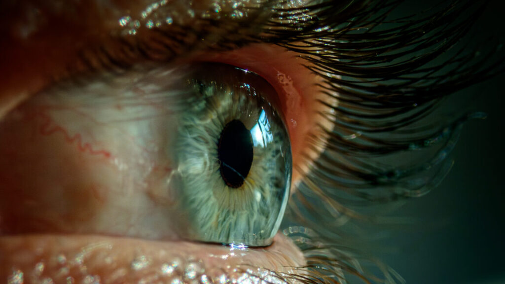
For people with OSA, researchers speculate that the sleep disorder leads to hypoxic conditions (a lack of oxygen) in their tissue. This, in turn, could increase oxidative stress and reactive oxygen species in the tissue, which can spur the production of enzymes that break down elastin in the eyelid. Thus, the eyelids become lax and limp, allowing them to get into weird positions (such as inside out) and leading to chronic irritation of the eye surface.

The good news is that most people with floppy eye syndrome can manage the condition with conservative measures, such as CPAP for those with OSA, as did the woman in New York. But some may end up needing corrective surgery.

Bad sleep made woman’s eyelids so floppy they flipped inside out, got stuck Read More »

this-may-be-the-grossest-eye-pic-ever—but-the-cause-is-what’s-truly-horrifying

This may be the grossest eye pic ever—but the cause is what’s truly horrifying

Savage microbe

Whatever was laying waste to his eye seemed to have come from inside his own body, carried in his bloodstream—possibly the same thing that could explain the liver mass, lung nodules, and brain lesions. There was one explanation that fit the condition perfectly: hypervirulent Klebsiella pneumoniae or hvKP.

Classical K. pneumoniae is a germ that dwells in people’s intestinal tracts and is one that’s familiar to doctors. It’s known for lurking in health care settings and infecting vulnerable patients, often causing pneumonia or urinary tract infections. But hvKP is very different. In comparison, it’s a beefed-up bacteria with a rage complex. It was first identified in the 1980s in Taiwan—not for stalking weak patients in the hospital but for devastating healthy people in normal community settings.

An infection with hvKP—even in otherwise healthy people—is marked by metastatic infection. That is, the bacteria spreads throughout the body, usually starting with the liver, where it creates a pus-filled abscess. It then goes on a trip through the bloodstream, invading the lungs, brain, soft tissue, skin, and the eye (endogenous endophthalmitis). Putting it all together, the man had a completely typical clinical case of an hvKP infection.

Still, definitively identifying hvKP is tricky. Mucus from the man’s respiratory tract grew a species of Klebsiella, but there’s not yet a solid diagnostic test to differentiate hvKP from the classical variety. Since 2024, researchers have worked out a strategy of using the presence of five different virulence genes found on plasmids (relatively small, circular pieces of DNA, separate from chromosomal DNA, that can replicate on their own and be shared among bacteria.) But the method isn’t perfect—some classical K. pneumoniae can also carry the five genes.

A string test performed on the rare growth of Klebsiella pneumoniae from the sputum culture shows a positive result, with the formation of a viscous string with a height of greater than 5 mm.

A string test performed on the rare growth of Klebsiella pneumoniae from the sputum culture shows a positive result, with the formation of a viscous string with a height of greater than 5 mm. Credit: NEJM 2026

Another much simpler method is the string test, in which clinicians basically test the goopy-ness of the bacteria—hvKP is known for being sticky. For this test, a clinician grows the bacteria into a colony on a petri dish, then touches an inoculation loop to the colony and pulls up. If the string of attached goo stretches more than 5 mm off the petri dish, it’s considered positive for hvKP. This is (obviously) not a precise test.

This may be the grossest eye pic ever—but the cause is what’s truly horrifying Read More »

this-may-be-the-grossest-eye-pic-ever—but-the-cause-is-what’s-truly-horrifying

This may be the grossest eye pic ever—but the cause is what’s truly horrifying

Savage microbe

Whatever was laying waste to his eye seemed to have come from inside his own body, carried in his bloodstream—possibly the same thing that could explain the liver mass, lung nodules, and brain lesions. There was one explanation that fit the condition perfectly: hypervirulent Klebsiella pneumoniae or hvKP.

Classical K. pneumoniae is a germ that dwells in people’s intestinal tracts and is one that’s familiar to doctors. It’s known for lurking in health care settings and infecting vulnerable patients, often causing pneumonia or urinary tract infections. But, hvKP is very different. In comparison, it’s a beefed-up bacteria with a rage complex. It was first identified in the 1980s in Taiwan—not for stalking weak patients in the hospital, but devastating healthy people in normal community settings.

An infection with hvKP—even in otherwise healthy people—is marked by metastatic infection. That is, the bacteria spread throughout the body, usually starting with the liver, where it creates a pus-filled abscess. Then it goes on a trip through the bloodstream, invading the lungs, brain, soft tissue, skin, and the eye (endogenous endophthalmitis). Putting it all together, the man had a completely typical clinical case of an hvKP infection.

Still, definitely identifying hvKP is tricky. Mucus from the man’s respiratory tract grew a species of Klebsiella, but there is not yet a solid diagnostic test to differentiate hvKP from the classical variety. Just since 2024, researchers have worked out a strategy of using the presence of five different virulence genes found on plasmids (relatively small, circular pieces of DNA, separate from chromosomal DNA, that can replicate on their own and be shared among bacteria.) But the method isn’t perfect—some classical K. pneumoniae can also carry the five genes.

A string test performed on the rare growth of Klebsiella pneumoniae from the sputum culture shows a positive result, with the formation of a viscous string with a height of greater than 5 mm.

A string test performed on the rare growth of Klebsiella pneumoniae from the sputum culture shows a positive result, with the formation of a viscous string with a height of greater than 5 mm. Credit: NEJM 2026

Another, much simpler method is the string test, in which clinicians basically test the goopy-ness of the bacteria—hvKP is known for being sticky. For this test, a clinician grows the bacteria into a colony on a petri dish then touches an inoculation loop to the colony and pulls up. If the string of attached goo stretches more than 5 mm off the petri dish, it’s considered positive for hvKP. But, this is (obviously) not a precise test.

This may be the grossest eye pic ever—but the cause is what’s truly horrifying Read More »

four-inch-worm-hatches-in-woman’s-forehead,-wriggles-to-her-eyelid

Four-inch worm hatches in woman’s forehead, wriggles to her eyelid

Creeping

For anyone enjoying—or at least trying to enjoy—Thanksgiving in America, you can be thankful that these worms are not present in the US; they are exclusive to the “Old World,” that is Europe, Africa, and Asia, according to the Centers for Disease Control and Prevention. They’re often found in the Mediterranean region, but reports in recent years have noted that they seem to be expanding into new areas of Europe—particularly eastward and northward. In a report earlier this year of cases in Estonia, researchers noted that it is also emerging in Lithuania, Latvia, and Finland.

Researchers attribute the worm’s creep to climate change and globalization. But in another report this year of a case in Austria (thought to be acquired while the patient was vacationing in Greece), researchers also raised the speculation that the worms may be adapting to use humans as a true host. Researchers in Serbia suggested this in a 2023 case report, in which an infection led to microfilariae in the patient’s blood. The researchers speculated that such cases, considered rare, could be increasing.

For now, people in America have less to worry about. D. repens has not been found in the US, but it does have some relatives here that occasionally show up in humans, including D. immitis, the cause of dog heartworm, and D. tenuis. The latter can cause similar cases to D. repens, with worms wandering under the skin, particularly around the eye. So far, this worm has mainly been found in raccoons in Florida.

For those who do find a worm noodling through their skin, the outlook is generally good. Treatment includes surgical removal of the worm, which largely takes care of the problem, as well as anti-parasitic or antibiotic drugs to be sure to stamp out the infection or any co-infections. In the woman’s case, her symptoms disappeared after doctors pulled the worm from her eyelid.

Four-inch worm hatches in woman’s forehead, wriggles to her eyelid Read More »

man-suffers-rare-bee-sting-directly-to-the-eyeball—it-didn’t-go-well

Man suffers rare bee sting directly to the eyeball—it didn’t go well

Nightmare fuel —

He did recover. No disturbing images in the article, but a link for those who dare.

Bees fly to their hive.

Enlarge / Bees fly to their hive.

In what may be the biological equivalent to getting struck by lightning, a very unlucky man in the Philadelphia area took a very rare bee sting directly to the eyeball—and things went badly from there.

As one might expect, the 55-year-old went to the emergency department, where doctors tried to extract the injurious insect’s stinger from the man’s right eye. But it soon became apparent that they didn’t get it all.

Two days after the bee attack, the man went to the Wills Eye Hospital with worsening vision and pain in the pierced eye. At that point, the vision in his right eye had deteriorated to only being able to count fingers. The eye was swollen, inflamed, and bloodshot. Blood was visibly pooling at the bottom of his iris. And right at the border between the man’s cornea and the white of his eye, ophthalmologists spotted the problem: a teeny spear-like fragment of the bee’s stinger still stuck in place.

(Images of the eye and stinger fragment are here for those who aren’t squeamish. The white arrow in Panel A shows the location of the stinger fragment while the asterisk marks the pooled blood.)

Get thee to an ophthalmologist

In a report published recently in the New England Journal of Medicine, treating ophthalmology experts Talia Shoshany and Zeba Syed made a critical recommendation: If you happen to be among the ill-fated few who are stung in the eye by a bee, you should make sure to see an eye doctor specifically.

“I am not surprised that the ER missed a small fragment,” Shoshany told Ars over email. “They pulled out the majority of the stinger, but the small fragment was only able to be visualized at a slit lamp,” she said, referring to a microscope with a bright light used in eye exams. In this case, they visualized the stinger at 10X or 16X magnification with the additional help of a fluorescent dye. Moreover, after spotting it, the stinger fragment “needed to be pulled out with ophthalmic-specific micro-forceps.”

After finally getting the entirety of the wee dagger out, Shoshany and Syed prescribed a topical antibacterial and prednisolone eye drops (a steroid for inflammation). At a five-month follow-up, the patient had recovered and the vision in his right eye had improved to 20/25.

For those now in fear of eye stings, Soshany has some comforting words: “Ocular bee stings are very rare.” She noted this was the first one she had seen in her career. Although there are documented cases in the scientific literature, the incidence rate is unknown. The odds of getting struck by lightning, meanwhile, are 1 in 15,300, according to the National Weather Service.

But one troubling aspect of this case is that it’s unclear why the man was stung to begin with. According to Shoshany, the man worked on a property with a beehive, but he didn’t work with the insects himself. “He reports he was just walking by and several bees flew up to him; one stung him in the eye,” she said. It’s unclear what provoked them.

Man suffers rare bee sting directly to the eyeball—it didn’t go well Read More »