Biology

the-evolution-of-rationality:-how-chimps-process-conflicting-evidence

The evolution of rationality: How chimps process conflicting evidence

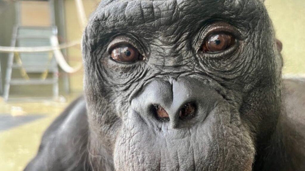
In the first step, the chimps got the auditory evidence, the same rattling sound coming from the first container. Then, they received indirect visual evidence: a trail of peanuts leading to the second container. At this point, the chimpanzees picked the first container, presumably because they viewed the auditory evidence as stronger. But then the team would remove a rock from the first container. The piece of rock suggested that it was not food that was making the rattling sound. “At this point, a rational agent should conclude, ‘The evidence I followed is now defeated and I should go for the other option,’” Engelmann told Ars. “And that’s exactly what the chimpanzees did.”

The team had 20 chimpanzees participating in all five experiments, and they followed the evidence significantly above chance level—in about 80 percent of the cases. “At the individual level, about 18 out of 20 chimpanzees followed this expected pattern,” Engelmann claims.

He views this study as one of the first steps to learn how rationality evolved and when the first sparks of rational thought appeared in nature. “We’re doing a lot of research to answer exactly this question,” Engelmann says.

The team thinks rationality is not an on/off switch; instead, different animals have different levels of rationality. “The first two experiments demonstrate a rudimentary form of rationality,” Engelmann says. “But experiments four and five are quite difficult and show a more advanced form of reflective rationality I expect only chimps and maybe bonobos to have.”

In his view, though, humans are still at least one level above the chimps. “Many people say reflective rationality is the final stage, but I think you can go even further. What humans have is something I would call social rationality,” Engelmann claims. “We can discuss and comment on each other’s thinking and in that process make each other even more rational.”

Sometimes, at least in humans, social interactions can also increase our irrationality instead. But chimps don’t seem to have this problem. Engelmann’s team is currently running a study focused on whether the choices chimps make are influenced by the choices of their fellow chimps. “The chimps only followed the other chimp’s decision when the other chimp had better evidence,” Engelmann says. “In this sense, chimps seem to be more rational than humans.”

Science, 2025. DOI: 10.1126/science.aeb7565

The evolution of rationality: How chimps process conflicting evidence Read More »

wyoming-dinosaur-mummies-give-us-a-new-view-of-duck-billed-species

Wyoming dinosaur mummies give us a new view of duck-billed species


Exquisitely preserved fossils come from a single site in Wyoming.

The scaly skin of a crest over the back of the juvenile duck-billed dinosaur Edmontosaurus annectens. Credit: Tyler Keillor/Fossil Lab

Edmontosaurus annectens, a large herbivore duck-billed dinosaur that lived toward the end of the Cretaceous period, was discovered back in 1908 in east-central Wyoming by C.H. Sternberg, a fossil collector. The skeleton, later housed at the American Museum of Natural History in New York and nicknamed the “AMNH mummy,” was covered by scaly skin imprinted in the surrounding sediment that gave us the first approximate idea of what the animal looked like.

More than a century later, a team of paleontologists led by Paul C. Sereno, a professor of organismal biology at the University of Chicago, got back to the same exact place where Sternberg dug up the first Edmontosaurus specimen. The researchers found two more Edmontosaurus mummies with all fleshy external anatomy imprinted in a sub-millimeter layer of clay. For the first time, we uncovered an accurate image of what Edmontosaurus really looked like, down to the tiniest details, like the size of its scales and the arrangement of spikes on its tail. And we were in for at least a few surprises.

Evolving images

Our view of Edmontosaurus changed over time, even before Sereno’s study. The initial drawing of Edmontosaurus was made in 1909 by Charles R. Knight, a famous paleoartist, who based his visualization on the first specimen found by Sternberg. “He was accurate in some ways, but he made a mistake in that he drew the crest extending throughout the entire length of the body,” Sereno says. The mummy Knight based his drawing on had no tail, so understandably, the artist used his imagination to fill in the gaps and made the Edmontosaurus look a little bit like a dragon.

An update to Knight’s image came in 1984 due to Jack Horner, one of the most influential American paleontologists, who found a section of Edmontosaurus tail that had spikes instead of a crest. “The specimen was not prepared very accurately, so he thought the spikes were rectangular and didn’t touch each other,” Sereno explains. “In his reconstruction he extended the spikes from the tail all the way to the head—which was wrong,” Sereno says. Over time, we ended up with many different, competing visions of Edmontosaurus. “But I think now we finally nailed down the way it truly looked,” Sereno claims.

To nail it down, Sereno’s team retraced the route to where Sternberg found the first Edmontosaurus mummy. This was not easy, because the team had to rely on Sternberg’s notes, which often referred to towns and villages that were no longer on the map. But based on interviews with Wyoming farmers, Sereno managed to reach the “mummy zone,” an area less than 10 kilometers in diameter, surprisingly abundant in Cretaceous fossils.

“To find dinosaurs, you need to understand geology,” Sereno says. And in the “mummy zone,” geological processes created something really special.

Dinosaur templating

The fossils are found in part of the Lance Formation, a geological formation that originated in the last three or so million years of the Cretaceous period, just before the dinosaurs’ extinction. It extends through North Dakota, South Dakota, Wyoming, Montana, and even to parts of Canada. “The formation is roughly 200 meters thick. But when you approach the mummy zone—surprise! The formation suddenly goes up to a thousand meters thick,” Sereno says. “The sedimentation rate in there was very high for some reason.”

Sereno thinks the most likely reason behind the high sedimentation rate was frequent and regular flooding of the area by a nearby river. These floods often drowned the unfortunate dinosaurs that roamed there and covered their bodies with mud and clay that congealed against a biofilm which formed at the surface of decaying carcasses. “It’s called clay templating, where the clay sticks to the outside of the skin and preserves a very thin layer, a mask, showing how the animal looked like,” Sereno says.

Clay templating is a process well-known by scientists studying deep-sea invertebrate organisms because that’s the only way they can be preserved. “It’s just no one ever thought it could happen to a large dinosaur buried in a river,” Sereno says. But it’s the best explanation for the Wyoming mummy zone, where Sereno’s team managed to retrieve two more Edmontosaurus skeletons surrounded by clay masks under 1 millimeter thick. These revealed the animal’s appearance with amazing, life-like accuracy.

As a result, the Edmontosaurus image got updated one more time. And some of the updates were rather striking.

Delicate elephants

Sereno’s team analyzed the newly discovered Edmontosaurus mummies with a barrage of modern imaging techniques like CT scans, X-rays, photogrammetry, and more. “We created a detailed model of the skin and wrapped it around the skeleton—some of these technologies were not even available 10 years ago,” Sereno says. The result was an updated Edmontosaurus image that includes changes to the crest, the spikes, and the appearance of its skin. Perhaps most surprisingly, it adds hooves to its legs.

It turned out both Knight and Horner were partially right about the look of Edmontosaurus’ back. The fleshy crest, as depicted by Knight, indeed started at the top of the head and extended rearward along the spine. The difference was that there was a point where this crest changed into a row of spikes, as depicted in the Horner version. The spikes were similar to the ones found on modern chameleons, where each spike corresponds one-to-one with the vertebrae underneath it.

“Another thing that was stunning in Edmontosaurus was the small size of its scales,” Sereno says. Most of the scales were just 1 to 4 millimeters across. They grew slightly larger toward the bottom of the tail, but even there they did not exceed 1 centimeter. “You can find such scales on a lizard, and we’re talking about an animal the size of an elephant,” Sereno adds. The skin covered with these super-tiny scales was also incredibly thin, which the team deduced from the wrinkles they found in their imagery.

And then came the hooves. “In a hoof, the nail goes around the toe and wraps, wedge-shaped, around its bottom,” Sereno explains. The Edmontosaurus had singular, central hooves on its fore legs with a “frog,” a triangular, rubbery structure at the underside. “They looked very much like equine hooves, so apparently these were not invented by mammals,” Sereno says. “Dinosaurs had them.” The hind legs that supported most of the animal’s weight, on the other hand, had three wedge-shaped hooves wrapped around three digits and a fleshy heel toward the back—a structure found in modern-day rhinos.

“There are so many amazing ‘firsts’ preserved in these duck-billed mummies,” Sereno says. “The earliest hooves were documented in a land vertebrate, the first confirmed hooved reptile, and the first hooved four-legged animal with different forelimb and hindlimb posture.” But Edmontosaurus, while first in many aspects, was not the last species Sereno’s team found in the mummy zone.

Looking for wild things

“When I was walking through the grass in the mummy zone for the first time, the first hill I found a T. rex in a concretion. Another mummy we found was a Triceratops,” Sereno says. Both these mummies are currently being examined and will be covered in the upcoming papers published by Sereno’s team. And both are unique in their own way.

The T. rex mummy was preserved in a surprisingly life-like pose, which Sereno thinks indicates the predator might have been buried alive. Edmontosaurus mummies, on the other hand, were positioned in a death pose, which meant the animals most likely died up to a week before the mud covered their carcasses. This, in principle, should make the T. rex clay mask even more true-to-life, since there should be no need to account for desiccation and decay when reconstructing the animal’s image.

Sereno, though, seems to be even more excited about the Triceratops mummy. “We already found Triceratops scales were 10 times larger than the largest scales on the Edmontosaurus, and its skin had no wrinkles, so it was significantly thicker. And we’re talking about animals of similar size living in the same area and in the same time,” Sereno says. To him, this could indicate that the physiology of the Triceratops and Edmontosaurus was radically different.

“We are in the age of discovery. There are so many things to come. It’s just the beginning,” Sereno says. “Anyway, the next two mummies we want to cover are the Triceratops and the T. Rex. And I can already tell you what we have with the Triceratops is wild,” he adds.

Science, 2025. DOI: 10.1126/science.adw3536

Photo of Jacek Krywko

Jacek Krywko is a freelance science and technology writer who covers space exploration, artificial intelligence research, computer science, and all sorts of engineering wizardry.

Wyoming dinosaur mummies give us a new view of duck-billed species Read More »

world’s-oldest-rna-extracted-from-ice-age-woolly-mammoth

World’s oldest RNA extracted from ice age woolly mammoth

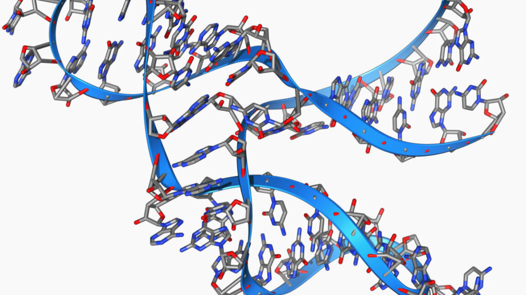
A young woolly mammoth now known as Yuka was frozen in the Siberian permafrost for about 40,000 years before it was discovered by local tusk hunters in 2010. The hunters soon handed it over to scientists, who were excited to see its exquisite level of preservation, with skin, muscle tissue, and even reddish hair intact. Later research showed that, while full cloning was impossible, Yuka’s DNA was in such good condition that some cell nuclei could even begin limited activity when placed inside mouse eggs.

Now, a team has successfully sequenced Yuka’s RNA—a feat many researchers once thought impossible. Researchers at Stockholm University carefully ground up bits of muscle and other tissue from Yuka and nine other woolly mammoths, then used special chemical treatments to pull out any remaining RNA fragments, which are normally thought to be much too fragile to survive even a few hours after an organism has died. Scientists go to great lengths to extract RNA even from fresh samples, and most previous attempts with very old specimens have either failed or been contaminated.

A different view

The team used RNA-handling methods adapted for ancient, fragmented molecules. Their scientific séance allowed them to explore information that had never been accessible before, including which genes were active when Yuka died. In the creature’s final panicked moments, its muscles were tensing and its cells were signaling distress—perhaps unsurprising since Yuka is thought to have died as a result of a cave lion attack.

It’s an exquisite level of detail, and one that scientists can’t get from just analyzing DNA. “With RNA, you can access the actual biology of the cell or tissue happening in real time within the last moments of life of the organism,” said Emilio Mármol, a researcher who led the study. “In simple terms, studying DNA alone can give you lots of information about the whole evolutionary history and ancestry of the organism under study. “Obtaining this fragile and mostly forgotten layer of the cell biology in old tissues/specimens, you can get for the first time a full picture of the whole pipeline of life (from DNA to proteins, with RNA as an intermediate messenger).”

World’s oldest RNA extracted from ice age woolly mammoth Read More »

corals-survived-past-climate-changes-by-retreating-to-the-deeps

Corals survived past climate changes by retreating to the deeps


A recent die-off in Florida puts the spotlight on corals’ survival strategies.

Scientists have found that the 2023 marine heat wave caused “functional extinction” of two Acropora reef-building coral species living in the Florida Reef, which stretches from the Dry Tortugas National Park to Miami.

“At this point, we do not think there’s much of a chance for natural recovery—their numbers are so low that successful reproduction is incredibly unlikely,” said Ross Cunning, a coral biologist at the John G. Shedd Aquarium.

This isn’t the first time corals have faced the borderline of extinction over the last 460 million years, and they have always managed to bounce back and recolonize habitats lost during severe climate changes. The problem is that we won’t live long enough to see them doing that again.

Killer heat waves

Marine heat waves kill corals by messing with the photosynthetic machinery of symbiotic microalgae that live in the corals’ tissues. When the temperature of water goes up too much, the microalgae start producing reactive oxygen species instead of nutritious sugars. The reactive oxygen is toxic to corals, which respond by expelling the microalgae. This solves the toxicity problem, but it also starves the corals and causes them to bleach (the algae are the source of their yellowish color).

The 2023 marine heat wave was not the first to hit the Florida Reef—it was the ninth on record. “Those eight previous heat waves also had major negative effects on coral reefs, causing widespread mortality,” Cunning told Ars. “But the 2023 heat wave blew all other heat waves out of the water. It was 2.2 to four times greater in magnitude than anything that came before it.”

Cunning’s team monitored two Acropora coral species: the staghorn and elkhorn. “They are both branching corals,” Cunning explained. “The staghorn has pointy branches that form dense thickets, whereas elkhorn produces arm-like branches that reach up and grow toward the surface, producing highly complex three dimensionality, like a canopy in the forest.”

He and his colleagues chose those two species because they essentially built the Florida Reef. They also grow the fastest among all Florida Reef corals, which means they are essential for its ability to recover from damage. “Acropora corals were the primary reef builders for the last ten thousand years,” Cunning said. Unfortunately, they also showed the highest levels of mortality due to heat waves.

Coral apocalypse

Cunning’s team found the mortality rate among Acropora corals reached 100 percent in the Dry Tortugas National Park, which is at the southernmost end of the Florida Reef. Moving north to Lower Keys, Middle Keys, and most of the Upper Keys, the mortality stayed at between 98 and 100 percent.

“Once you start moving a little bit further north, there’s the Biscayne National Park, where mortality rates were at 90 percent,” Cunning said. “It wasn’t until the furthest northern extent of the reef in Miami and Broward counties where mortality dropped to just 38 percent thanks to cooler temperatures that occurred there.”

Still, the mortality rate was exceptionally high throughout most of Acropora colonies across the Florida Reef. “What we’re facing is a functional extinction,” Cunning said.

But corals have been around for about 460 million years, and they have survived multiple mass extinction events, including the one that wiped out the dinosaurs. As vulnerable as they appear, corals seemingly have some get-out-of-death card they always pull when things turn really bad for them. This card, most likely, is buried deep in their genome.

Ancestral strength

“There have been studies looking into the evolutionary history of corals, but the difference between those and our work lies in technology,” said Claudia Francesca Vaga, a marine biologist at the Smithsonian Institution.

Her team looked at ultra conserved elements, stretches of DNA that are nearly identical across even distantly related species. These elements were used to build the most extensive phylogenetic tree of corals to date. Based on the genomic data and fossil evidence, Vaga’s team analyzed how 274 stony coral species are related to one another to retrace their common ancestor and reconstruct how they evolved from it.

“We managed to confirm that the first common ancestor of stony corals was most likely solitary—it didn’t live in colonies, and it didn’t have symbionts,” Vaga said.

The very first coral most likely did not rely on algae to produce its nutrients, which means it was immune to bleaching. It was also not attached to a substrate, so it could move from one habitat to another. Another advantage the first corals had was that they were not particularly picky—they could live just as well in the shallow waters as in the deep sea, since they didn’t get most of their nutrients from their photosynthetic symbionts.

Descending from these incredibly resilient ancestors, corals started to specialize. “We learned that symbiosis and coloniality can be acquired independently by stony coral linages and that it happened multiple times,” Vaga said.

Based on her team’s research, past mass extinction events usually wiped out 90 percent of the species living in shallow waters—the ones that were colonial and reliant on symbionts. “But each such extinction triggered a process of retaking the shallows by the more resilient deep-sea corals, which in time evolved symbiosis and coloniality again,” Vaga said.

Thanks to corals’ deep-sea cousins, even the most extreme environmental changes—global warming or sudden, severe variations in the oceans’ acidity or oxygen levels—could not kill them for good. Each mass extinction event they’ve been through just reverted them to factory settings and made them start over from scratch.

The only catch here is time. “We’re talking about four to five million years before coral populations recover,” Vaga said.

Long way back

According to Cunning, the consequence of Acropora corals’ extinction in the Florida Reef is a lower overall reef-building rate, which will lead to reduced biodiversity in the reef’s ecosystem. “There are going to be cascading effects, and humans will be impacted as well. Reefs protect our coastlines by buffering over 90 percent of wave energy,” Cunning said.

In Florida, where coastlines are heavily urbanized, this may translate into hundreds of millions of dollars per year in damages.

But Cunning said we still have means at our disposal to save Acropora corals. “We’re not going to give up on them,” he said.

One option for improving the resilience of corals could be to crossbreed them with species from outside of Florida Reef, ideally ones that live in warmer places and are better adapted to heat. “The first tests of this approach are underway right now in Florida; elkhorn corals were cross bred between Florida parents and Honduran parents,” Cunning said. He hopes this will help produce a new generation of corals that has a better shot at surviving the next heat wave.

Other interventions include manipulating corals’ algal symbionts. “There are many different species of algae with different levels of heat tolerance,” Cunning said. To him, a possible way forward would be to pair the Acropora corals with more heat-tolerant symbionts. “This should alter the bleaching threshold in these corals,” he explained.

Still, even interventions like these will take a very long time to make a difference. “But if four or five million years is the benchmark to beat, then yeah, it’s hopefully going to happen faster than that,” Cunning said.

The upside is that corals will likely pull off their de-extinction trick once again, even if we do absolutely nothing to help them. “In a few million years, they will redevelop coloniality, redevelop symbiosis, and rebuild something similar to the coral reefs we have today,” Vaga said. “This is good news for them. Not necessarily for us.”

Science, 2025.  DOI: 10.1126/science.adx7825

Nature, 2025.  DOI: 10.1038/s41586-025-09615-6

Photo of Jacek Krywko

Jacek Krywko is a freelance science and technology writer who covers space exploration, artificial intelligence research, computer science, and all sorts of engineering wizardry.

Corals survived past climate changes by retreating to the deeps Read More »

some-stinkbugs’-legs-carry-a-mobile-fungal-garden

Some stinkbugs’ legs carry a mobile fungal garden

Many insect species hear using tympanal organs, membranes roughly resembling our eardrums but located on their legs. Grasshoppers, mantises, and moths all have them, and for decades, we thought that female stinkbugs of the Dinidoridae family have them, too, although located a bit unusually on their hind rather than front legs.

Suspecting that they use their hind leg tympanal organs to listen to male courtship songs, a team of Japanese researchers took a closer look at the organs in Megymenum gracilicorne, a Dinidoridae stinkbug species native to Japan. They discovered that these “tympanal organs” were not what they seemed. They’re actually mobile fungal nurseries of a kind we’ve never seen before.

Portable gardens

Dinidoridae is a small stinkbug family that lives exclusively in Asia. The bug did attract some scientific attention, but not nearly as much as its larger relatives like Pentatomidae. Prior work looking specifically into organs growing on the hind legs of Dinidoridae females was thus somewhat limited. “Most research relied on taxonomic and morphological approaches. Some taxonomists did describe that female Dinidoridae stinkbugs have an enlarged part on the hind legs that looks like the tympanal organ you can find, for example, in crickets,” said Takema Fukatsu, an evolutionary biologist at the National Institute of Advanced Industrial Science and Technology in Tokyo.

Based on that appearance, these parts were classified as tympanal organs—the case was closed, and it stayed closed until Fukatsu’s team started examining them more closely. Most insects have tympanal organs on their front legs, not hind legs, or on abdominal segments. The initial goal of Fukatsu’s study was to figure out what impact this unusual position has on Dinidoridae females’ ability to hear sounds.

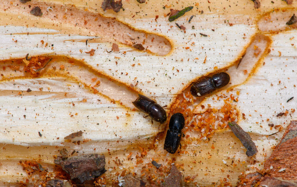
Early on in the study, it turned out that whatever Dinidoridae females have on their hind legs, they are not tympanal organs. “We found no tympanal membrane and no sensory neurons, so the enlarged parts on the hind legs had nothing to do with hearing,” Fukatsu explained. Instead, the organ had thousands of small pores filled with benign filamentous fungi. The pores were connected to secretory cells that released substances that Fukatsu’s team hypothesized were nutrients enabling the fungi to grow.

Some stinkbugs’ legs carry a mobile fungal garden Read More »

neural-network-finds-an-enzyme-that-can-break-down-polyurethane

Neural network finds an enzyme that can break down polyurethane

You’ll often hear plastic pollution referred to as a problem. But the reality is that it’s multiple problems. Depending on the properties we need, we form plastics out of different polymers, each of which is held together by a distinct type of chemical bond. So the method we use to break down one type of polymer may be incompatible with the chemistry of another.

That problem is why, even though we’ve had success finding enzymes that break down common plastics like polyesters and PET, they’re only partial solutions to plastic waste. However, researchers aren’t sitting back and basking in the triumph of partial solutions, and they’ve now got very sophisticated protein design tools to help them out.

That’s the story behind a completely new enzyme that researchers developed to break down polyurethane, the polymer commonly used to make foam cushioning, among other things. The new enzyme is compatible with an industrial-style recycling process that breaks the polymer down into its basic building blocks, which can be used to form fresh polyurethane.

Breaking down polyurethane

Image of a set of chemical bonds. From left to right there is an X, then a single bond to an oxygen, then a single bond to an oxygen that's double-bonded to carbon, then a single bond to a nitrogen, then a single bond to another X.

The basics of the chemical bonds that link polyurethanes. The rest of the polymer is represented by X’s here.

The new paper that describes the development of this enzyme lays out the scale of the problem: In 2024, we made 22 million metric tons of polyurethane. The urethane bond that defines these involves a nitrogen bonded to a carbon that in turn is bonded to two oxygens, one of which links into the rest of the polymer. The rest of the polymer, linked by these bonds, can be fairly complex and often contains ringed structures related to benzene.

Digesting polyurethanes is challenging. Individual polymer chains are often extensively cross-linked, and the bulky structures can make it difficult for enzymes to get at the bonds they can digest. A chemical called diethylene glycol can partially break these molecules down, but only at elevated temperatures. And it leaves behind a complicated mess of chemicals that can’t be fed back into any useful reactions. Instead, it’s typically incinerated as hazardous waste.

Neural network finds an enzyme that can break down polyurethane Read More »

why-imperfection-could-be-key-to-turing-patterns-in-nature

Why imperfection could be key to Turing patterns in nature

In essence, it’s a type of symmetry breaking. Any two processes that act as activator and inhibitor will produce periodic patterns and can be modeled using Turing’s diffusion function. The challenge is moving from Turing’s admittedly simplified model to pinpointing the precise mechanisms serving in the activator and inhibitor roles.

This is especially challenging in biology. Per the authors of this latest paper, the classical approach to a Turing mechanism balances reaction and diffusion using a single length scale, but biological patterns often incorporate multiscale structures, grain-like textures, or certain inherent imperfections. And the resulting patterns are often much blurrier than those found in nature.

Can you say “diffusiopherosis”?

Simulated hexagon and stripe patterns obtained by diffusiophoretic assembly of two types of cells on top of the chemical patterns. Credit: Siamak Mirfendereski and Ankur Gupta/CU Boulder

In 2023, UCB biochemical engineers Ankur Gupta and Benjamin Alessio developed a new model that added diffusiopherosis into the mix. It’s a process by which colloids are transported via differences in solute concentration gradients—the same process by which soap diffuses out of laundry in water, dragging particles of dirt out of the fabric. Gupta and Alessio successfully used their new model to simulate the distinctive hexagon pattern (alternating purple and black) on the ornate boxfish, native to Australia, achieving much sharper outlines than the model originally proposed by Turing.

The problem was that the simulations produced patterns that were too perfect: hexagons that were all the same size and shape and an identical distance apart. Animal patterns in nature, by contrast, are never perfectly uniform. So Gupta and his UCB co-author on this latest paper, Siamak Mirfendereski, figured out how to tweak the model to get the pattern outputs they desired. All they had to do was define specific sizes for individual cells. For instance, larger cells create thicker outlines, and when they cluster, they produce broader patterns. And sometimes the cells jam up and break up a stripe. Their revised simulations produced patterns and textures very similar to those found in nature.

“Imperfections are everywhere in nature,” said Gupta. “We proposed a simple idea that can explain how cells assemble to create these variations. We are drawing inspiration from the imperfect beauty of [a] natural system and hope to harness these imperfections for new kinds of functionality in the future.” Possible future applications include “smart” camouflage fabrics that can change color to better blend with the surrounding environment, or more effective targeted drug delivery systems.

Matter, 2025. DOI: 10.1016/j.matt.2025.102513 (About DOIs).

Why imperfection could be key to Turing patterns in nature Read More »

clinical-trial-of-a-technique-that-could-give-everyone-the-best-antibodies

Clinical trial of a technique that could give everyone the best antibodies


If we ID the DNA for a great antibody, anyone can now make it.

One of the things that emerging diseases, including the COVID and Zika pandemics, have taught us is that it’s tough to keep up with infectious diseases in the modern world. Things like air travel can allow a virus to spread faster than our ability to develop therapies. But that doesn’t mean biotech has stood still; companies have been developing technologies that could allow us to rapidly respond to future threats.

There are a lot of ideas out there. But this week saw some early clinical trial results of one technique that could be useful for a range of infectious diseases. We’ll go over the results as a way to illustrate the sort of thinking that’s going on, along with the technologies we have available to pursue the resulting ideas.

The best antibodies

Any emerging disease leaves a mass of antibodies in its wake—those made by people in response to infections and vaccines, those made by lab animals we use to study the infectious agent, and so on. Some of these only have a weak affinity for the disease-causing agent, but some of them turn out to be what are called “broadly neutralizing.” These stick with high affinity not only to the original pathogen, but most or all of its variants, and possibly some related viruses.

Once an antibody latches on to a pathogen, broadly neutralizing antibodies inactivate it (as their name implies). This is typically because these antibodies bind to a site that’s necessary for a protein’s function. For example, broadly neutralizing antibodies to HIV bind to the proteins that help this virus enter immune cells.

Unfortunately, not everyone develops broadly neutralizing antibodies, and certainly doesn’t do so in time to prevent infections. And we haven’t figured out a way of designing vaccinations that ensure their generation. So we’re often found ourselves stuck with knowing what antibodies we’d like to see people making while having no way of ensuring that they do.

One of the options we’ve developed is to just mass-produce broadly neutralizing antibodies and inject them into people. This has been approved for use against Ebola and provided an early treatment during the COVID pandemic. This approach has some practical limitations, though. For starters, the antibodies have a finite life span in the bloodstream, so injections may need to be repeated. In addition, making and purifying enough antibodies in bulk isn’t the easiest thing in the world, and they generally need to be kept refrigerated during the distribution, limiting the areas where they can be used.

So, a number of companies have been looking at an alternative: getting people to make their own. This could potentially lead to longer-lived protection, even ensuring the antibodies are present to block future infections if the DNA survives long enough.

Genes and volts

Once you identify cells that produce broadly neutralizing antibodies, it’s relatively simple to clone those genes and put them into a chunk of DNA that will ensure that they’ll be produced by any human cell. If we could get that DNA into a person’s cells, broadly neutralizing antibodies are the result. And a number of approaches have been tried to handle that “if.” Most of them have inserted the genes needed to make the antibodies into a harmless, non-infectious virus, and then injected that virus into volunteers. Unfortunately, these viruses have tended to set off a separate immune response, which causes more significant side effects and may limit how often this approach can be used.

This brings us to the technique being used here. In this case, the researchers placed the antibody genes in a circular loop of DNA called a plasmid. This is enough to ensure that the DNA doesn’t get digested immediately and to get the antibody genes made into proteins. But it does nothing to help get the DNA inside of cells.

The research team, a mixture of people from a biotech company and academic labs, used a commercial injection setup that mixes the injection of the DNA with short pulses of electricity. The electricity disrupts the cell membrane, allowing the plasmid DNA to make it inside cells. Based on animal testing, doing this in muscle cells is enough to turn the muscles into factories producing lots of broadly neutralizing antibodies.

The new study was meant to test the safety of doing that in humans. The team recruited 44 participants, testing various doses of two antibody-producing plasmids and injection schedules. All but four of the subjects completed the study; three of those who dropped out had all been testing a routine with the electric pulses happening very quickly, which turned out to be unpleasant. Fortunately, it didn’t seem to make any difference to the production of antibodies.

While there were a lot of adverse reactions, most of these were associated with the injection itself: muscle pain at the site, a scab forming afterward, and a reddening of the skin. The worst problem appeared to be a single case of moderate muscle pain that persisted for a couple of days.

In all but one volunteer, the injection resulted in stable production of the two antibodies for at least 72 weeks following the injection; the single exception only made one of the two. That’s “at least” 72 weeks because that’s when they stopped testing—there was no indication that levels were dropping at this point. Injecting more DNA led to more variability in the amount of antibody produced, but that amount quickly maxed out. More total injections also boosted the level of antibody production. But even the minimal procedure—two injections of the lowest concentration tested—resulted in significant and stable antibodies.

And, as expected, these antibodies blocked the virus they were directed against: SARS-CoV-2.

The caveats

This approach seems to work—we can seemingly get anybody to make broadly neutralizing antibodies for months at a time. What’s the hitch? For starters, this isn’t necessarily great for a rapidly emerging pandemic. It takes a while to identify broadly neutralizing antibodies after a pathogen is identified. And, while it’s simple to ship DNA around the world to where it will be needed, injection setups that also produce the small electric pulses are not exactly standard equipment even in industrialized countries, much less the Global South.

Then there’s the issue of whether this really is a longer-term fix. Widespread use of broadly neutralizing antibodies will create a strong selective pressure for the evolution of variants that the antibody can no longer bind to. That may not always be a problem—broadly neutralizing antibodies generally bind to parts of proteins that are absolutely essential for the proteins’ function, and so it may not be possible to change those while maintaining the function. But that’s unlikely to always be the case.

In the end, however, social acceptance may end up being the biggest problem. People had an utter freakout over unfounded conspiracies that the RNA of COVID vaccines would somehow lead to permanent genetic changes. Presumably, having DNA that’s stable for months would be even harder for some segments of the public to swallow.

Nature Medicine, 2025. DOI: 10.1038/s41591-025-03969-0 (About DOIs).

Photo of John Timmer

John is Ars Technica’s science editor. He has a Bachelor of Arts in Biochemistry from Columbia University, and a Ph.D. in Molecular and Cell Biology from the University of California, Berkeley. When physically separated from his keyboard, he tends to seek out a bicycle, or a scenic location for communing with his hiking boots.

Clinical trial of a technique that could give everyone the best antibodies Read More »

dinosaurs-may-have-flourished-right-up-to-when-the-asteroid-hit

Dinosaurs may have flourished right up to when the asteroid hit

That seemingly changes as of now, with new argon dating of strata from the Naashoibito Member in the San Juan Basin of present-day New Mexico. Many dinosaur fossils have been obtained from this region, and we know the site differs from the sort of ecosystem found at Hell Creek. But it was previously thought to date back closer to a million years before the mass extinction. The new dates, plus the alignment of magnetic field reversals, tell us that the ecosystem was a contemporary of the one in Hell Creek, and dates to the last few hundred thousand years prior to the mass extinction.

Diverse ecosystems

The fossils at Naashoibito have revealed an ecosystem we now label the “Alamo Wash local fauna.” And they’re fairly distinct from the ones found in Wyoming, despite being just 1,500 kilometers further south. Analyzing the species present using ecological measures, the researchers found that dinosaurs formed two “bioprovinces” in the late Cretaceous—essentially, there were distinct ecosystems present in the northern and southern areas.

This doesn’t seem to be an artifact of the sites, as mammalian fossils seem to reflect a single community across both areas near the mass extinction, but had distinct ecologies both earlier and after. The researchers propose that temperature differences were the key drivers of the distinction, something that may have had less of an impact on mammals, which are generally better at controlling their own temperatures.

Overall, the researchers conclude that, rather than being dominated by a small number of major species, “dinosaurs were thriving in New Mexico until the end of the Cretaceous.”

While this speaks directly to the idea that limited diversity may have primed the dinosaurs for extinction, it also may have implications for the impact of the contemporaneous eruptions in the Deccan Traps. If these were having a major global impact, then it’s a bit unlikely that dinosaurs would be thriving anywhere.

Even with the new data, however, our picture is still limited to the ecosystems present on the North American continent. We do have fossils from elsewhere, but they’re not exactly dated. There are some indications of dinosaurs in the late Cretaceous in Europe and South America, but we don’t have a clear picture of the ecosystems in which they were found. So, while these findings help clarify the diversity of dinosaurs in the time leading up to their extinction, there’s still a lot left to learn.

Science, 2025. DOI: 10.1126/science.adw3282 (About DOIs).

Dinosaurs may have flourished right up to when the asteroid hit Read More »

bats-eat-the-birds-they-pluck-from-the-sky-while-on-the-wing

Bats eat the birds they pluck from the sky while on the wing

There are three species of bats that eat birds. We know that because we have found feathers and other avian remains in their feces. What we didn’t know was how exactly they hunt birds, which are quite a bit heavier, faster, and stronger than the insects bats usually dine on.

To find out, Elena Tena, a biologist at Doñana Biological Station in Seville, Spain, and her colleagues attached ultra-light sensors to Nyctalus Iasiopterus, the largest bats in Europe. What they found was jaw-droppingly brutal.

Inconspicuous interceptors

Nyctalus Iasiopterus, otherwise known as greater noctule bats, have a wingspan of about 45 centimeters. They have reddish-brown or chestnut fur with a slightly paler underside, and usually weigh around 40 to 60 grams. Despite that minimal weight, they are the largest of the three bat species known to eat birds, so the key challenge in getting a glimpse into the way they hunt was finding sensors light enough to not impede the bats’ flight.

Cameras, which are the usual go-to sensor, were out of the question. “Bats hunt at night, so you’d need night vision cameras, which together with batteries are too heavy for a bat to carry. Our sensors had to weigh below 10 percent of the weight of the bat—four to six grams,” Tena explained.

Tena and her team explored several alternative approaches throughout the last decade, including watching the bats from the ground or using military-grade radars. But even then, catching the hunting bats red-handed remained impossible.

In recent years, the technology and miniaturization finally caught up with Tena’s needs, and the team found the right sensors for the job and attached them to 14 greater noctule bats over the course of two years. The tags used in the study weighed around four grams, could run for several hours, and registered sound, altitude, and acceleration. This gave Tena and her colleagues a detailed picture of the bats’ behavior in the night sky. The recordings included both ambient environmental sounds and the ultra-frequency bursts bats use for echolocation. Combining altitude with accelerometer readouts enabled scientists to trace the bats’ movements through all their fast-paced turns, dives, and maneuvers.

Bats eat the birds they pluck from the sky while on the wing Read More »

dna-analysis-reveals-likely-pathogens-that-killed-napoleon’s-army

DNA analysis reveals likely pathogens that killed Napoleon’s army

State-of-the-art methodologies

Painting of Napoleon's army.

Rascovan and his co-authors note in their paper that the 2006 study relied upon outdated PCR-based technologies for its DNA analysis. As for the virus family detected in the Kalingrad dental pulp, they argue that those viruses are both ubiquitous and usually asymptomatic in humans—and thus are unlikely to be the primary culprits for the diseases that wiped out the French army. So Rascovan’s team decided to use current state-of-the-art DNA methodologies to re-analyze a different set of remains of Napoleonic soldiers who died in Vilnius.

“In most ancient human remains, pathogen DNA is extremely fragmented and only present in very low quantities, which makes it very difficult to obtain whole genomes,” said Rascovan. “So we need methods capable of unambiguously identifying infectious agents from these weak signals, and sometimes even pinpointing lineages, to explore the pathogenic diversity of the past.”

An 1812 report from one of Napoleon’s physicians, J.R.L. de Kirckhoff, specifically noted typhus, dysentery, and diarrhea after the soldiers arrived in Vilnius, which he attributed to large barrels of salted beets the starving troops consumed, “greatly upsetting us and strongly irritating the intestinal tract.” Rascovan et al. note that such symptoms could accompany any number of conditions or diseases common to 19th-century Europe. “Even today, two centuries later, it would still be impossible to perform a differential diagnosis between typhus, typhoid, or paratyphoid fever based solely on the symptoms or the testimonies of survivors,” the authors wrote.

Imperial Guard button discovered during excavation

Imperial Guard button discovered during excavation. Credit: UMR 6578 Aix-Marseille Université, CNRS, EFS

Over 3,200 individual remains, almost all men between the ages of 20 and 50, were excavated from the mass grave at Vilnius. Rascovan et al. focused on 13 teeth from 13 different individuals. To compensate for the degraded nature of the 200-year-old genome fragments, co-authors at the University of Tartu in Estonia helped develop a multistep authentication method to more accurately identify pathogens in the samples. In some cases, they were even able to identify a specific lineage.

DNA analysis reveals likely pathogens that killed Napoleon’s army Read More »

even-with-protections,-wolves-still-fear-humans

Even with protections, wolves still fear humans

This quickly became an issue, at least for some people. Mieczysław Kacprzak, an MP from Poland’s PSL Party, currently in the ruling coalition, addressed the parliament in December 2017, saying that wolves were roaming suburban roads and streets, terrorizing citizens—in his view, a tragedy waiting to happen. He also said children were afraid to go to school because of wolves and asked for support from the Ministry of Agriculture, which could lift the ban on hunting. An article in “Łowczy Polski,” a journal of the Polish hunting community with a title that translates as “The Polish Huntsman,” later backed these pro-hunting arguments, claiming wolves were a threat to humans, especially children.

The idea was that wolves, in the absence of hunting, ceased to perceive humans as a threat and felt encouraged to approach them. But it was an idea that was largely supported by anecdote. “We found this was not the case,” says Liana Zanette, a biologist at Western University and co-author of the study.

Super predators

To figure out if wolves really were no longer afraid of humans, Zanette, Clinchy, and their colleagues set up 24 camera traps in the Tuchola Forest. “Our Polish colleagues and co-authors, especially Maciej Szewczyk, helped us set those traps in places where we were most likely to find wolves,” Zanette says. “Maciej was literally saying ‘pick this tree,’ or ‘this crossroads.’” When sensors in the traps detected an animal nearby, the system took a photo and played one of three sounds, chosen at random.

The first sound was chirping birds, which the team used as a control. “We chose birds because this is a typical part of forest soundscape and we assumed wolves would not find this threatening,” Clinchy says. The next sound was barking dogs. The team picked this one because a dog is another large carnivore living in the same ecosystem, so it was expected to scare wolves. The third sound was just people talking calmly in Polish. Zanette, Clinchy, and their colleagues quantified the level of fear each sound caused in wolves by measuring how quickly they vacated the area upon hearing it.

Even with protections, wolves still fear humans Read More »