Remembering where your meals came from key for a small bird’s survival
Where’d I leave that again? —
For small birds, remembering where the food is beats forgetting when it’s gone.

It seems like common sense that being smart should increase the chances of survival in wild animals. Yet for a long time, scientists couldn’t demonstrate that because it was unclear how to tell exactly if a lion or a crocodile or a mountain chickadee was actually smart or not. Our best shots, so far, were looking at indirect metrics like brain size or doing lab tests of various cognitive skills such as reversal learning, an ability that can help an animal adapt to a changing environment.
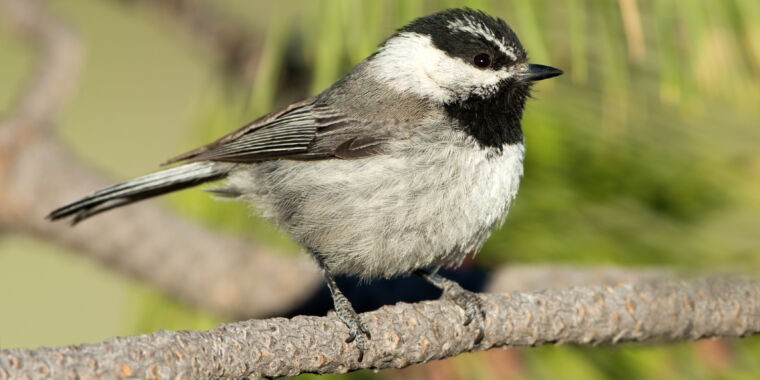
But a new, large-scale study on wild mountain chickadees, led by Joseph Welklin, an evolutionary biologist at the University of Nevada, showed that neither brain size nor reversal learning skills were correlated with survival. What mattered most for chickadees, small birds that save stashes of food, was simply remembering where they cached all their food. A chickadee didn’t need to be a genius to survive; it just needed to be good at its job.
Testing bird brains
“Chickadees cache one food item in one location, and they do this across a big area. They can have tens of thousands of caches. They do this in the fall and then, in the winter, they use a special kind of spatial memory to find those caches and retrieve the food. They are little birds, weight is like 12 grams, and they need to eat almost all the time. If they don’t eat for a few hours, they die,” explains Vladimir Pravosudov, an ornithologist at the University of Nevada and senior co-author of the study.
The team chose the chickadees to study the impact cognitive skills had on survival because the failure to find their caches was their most common cause of death. This way, the team hoped, the impact of other factors like predation or disease would be minimized.
First, however, Welklin and his colleagues had to come up with a way to test cognitive skills in a fairly large population of chickadees. They did it by placing a metal square with two smart feeders attached to each side among the trees where the chickadees lived. “The feeders were equipped with RFID receivers that recognized the signal whenever a chickadee, previously marked with a microchip-fitted leg band, landed near them and opened the doors to dispense a single seed,” Welklin says. After a few days spent getting the chickadees familiar with the door-opening mechanism, the team started running tests.
The first task was aimed at testing how good different chickadees were at their most important job: associating a location with food and remembering where it was. To this end, each of the 227 chickadees participating in the study was assigned just one feeder that opened when they landed on it; all the other feeders remained closed. A chickadee’s performance was measured by the number of trials it needed to figure out which feeder would serve it, and how many errors (landings on the wrong feeders) it made over four days. “If you were to find the right feeder at random, it should take you 3.5 trials on average. All the birds learned and performed way better than chance,” Pravosudov says.
The second task was meant to test reversal learning skills, widely considered the best predictor of survival. Once the chickadees learned the location of the reward-dispensing feeders, the locations were changed. The goal was to see how fast the birds would adapt to this change.
Once the results of both tests were in, the team monitored the birds using their microchip bands, catching them and changing the bands every year, for over six years. “Part of the reason that’s never been done in the past is just because it takes so much work,” says Welklin. But the work paid off in the end.
Remembering where your meals came from key for a small bird’s survival Read More »


