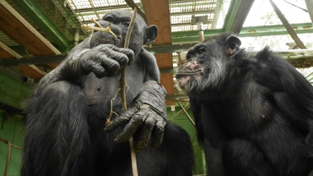
Biology

studies-reveal-new-clues-to-how-tardigrades-can-survive-intense-radiation

Studies reveal new clues to how tardigrades can survive intense radiation

It’s in the genes —

Radiation damages their DNA; they’re just able to repair that damage very quickly.

SEM Micrograph of a tardigrade, commonly known as a water bear

Enlarge / SEM Micrograph of a tardigrade, more commonly known as a “water bear” or “moss piglet.”

Cultura RM Exclusive/Gregory S. Paulson/Getty Images

Since the 1960s, scientists have known that the tiny tardigrade can withstand very intense radiation blasts 1,000 times stronger than what most other animals could endure. According to a new paper published in the journal Current Biology, it’s not that such ionizing radiation doesn’t damage tardigrades’ DNA; rather, the tardigrades are able to rapidly repair any such damage. The findings complement those of a separate study published in January that also explored tardigrades’ response to radiation.

“These animals are mounting an incredible response to radiation, and that seems to be a secret to their extreme survival abilities,” said co-author Courtney Clark-Hachtel, who was a postdoc in Bob Goldstein’s lab at the University of North Carolina at Chapel Hill, which has been conducting research into tardigrades for 25 years. “What we are learning about how tardigrades overcome radiation stress can lead to new ideas about how we might try to protect other animals and microorganisms from damaging radiation.”

As reported previously, tardigrades are micro-animals that can survive in the harshest conditions: extreme pressure, extreme temperature, radiation, dehydration, starvation—even exposure to the vacuum of outer space. The creatures were first described by German zoologist Johann Goeze in 1773. They were dubbed tardigrada (“slow steppers” or “slow walkers”) four years later by Lazzaro Spallanzani, an Italian biologist. That’s because tardigrades tend to lumber along like a bear. Since they can survive almost anywhere, they can be found in lots of places: deep-sea trenches, salt and freshwater sediments, tropical rain forests, the Antarctic, mud volcanoes, sand dunes, beaches, and lichen and moss. (Another name for them is “moss piglets.”)

When their moist habitat dries up, however, tardigrades go into a state known as “tun”—a kind of suspended animation, which the animals can remain in for as long as 10 years. When water begins to flow again, water bears absorb it to rehydrate and return to life. They’re not technically members of the extremophile class of organisms since they don’t so much thrive in extreme conditions as endure; technically, they belong to the class of extremotolerant organisms. But their hardiness makes tardigrades a favorite research subject for scientists.

For instance, a 2017 study demonstrated that tardigrades use a special kind of disordered protein to literally suspend their cells in a glass-like matrix that prevents damage. The researchers dubbed this a “tardigrade-specific intrinsically disordered protein” (TDP). In other words, the cells become vitrified. The more TDP genes a tardigrade species has, the more quickly and efficiently it goes into the tun state.

In 2021, another team of Japanese scientists called this “vitrification” hypothesis into question, citing experimental data suggesting that the 2017 findings could be attributed to water retention of the proteins. The following year, researchers at the University of Tokyo identified the mechanism to explain how tardigrades can survive extreme dehydration: cytoplasmic-abundant heat soluble (CAHS) proteins that form a protective gel-like network of filaments to protect dried-out cells. When the tardigrade rehydrates, the filaments gradually recede, ensuring that the cell isn’t stressed or damaged as it regains water.

When it comes to withstanding ionizing radiation, a 2016 study identified a DNA damage suppressor protein dubbed “Dsup” that seemed to shield tardigrade genes implanted into human cells from radiation damage. However, according to Clark-Hatchel et al., it still wasn’t clear whether this kind of protective mechanism was sufficient to fully account for tardigrades’ ability to withstand extreme radiation. Other species of tardigrade seem to lack Dsup proteins, yet still have the same high radiation tolerance, which suggests there could be other factors at play.

A team of French researchers at the French National Museum of Natural History in Paris ran a series of experiments in which they zapped water bear specimens with powerful gamma rays that would be lethal to humans. They published their results earlier this year in the journal eLife. The French team found that gamma rays did actually damage the tardigrade DNA, much like they would damage human cells. Since the tardigrades survived, this suggested the tardigrades were able to quickly repair the damaged DNA.

Further experiments with three different species (including one that lacks Dsup proteins) revealed the tardigrades were producing very high amounts of DNA repair proteins. They also found a similar uptick of proteins unique to tardigrades, most notably tardigrade DNA damage response protein 1 (TDR1), which seems to protect DNA from radiation. “We found that TDR1 protein interacts with DNA and forms aggregates at high concentration suggesting it may condensate DNA and act by preserving chromosome organization until DNA repair is accomplished,” the authors wrote.

Clark-Hatchel et al. independently arrived at similar conclusions from their own experiments. Taken together, the two studies confirm that this extremely rapid up-regulation of many DNA repair genes in response to exposure to ionizing radiation should be sufficient to explain the creatures’ impressive resistance to that radiation. It’s possible that there is a “synergy between protective and repair mechanisms” when it comes to tardigrade tolerance of ionizing radiation.

That said, “Why tardigrades have evolved a strong IR tolerance is enigmatic given that it is unlikely that tardigrades were exposed to high doses of ionizing radiation in their evolutionary history,” Clark-Hatchel et al. wrote.  They thought there could be a link to the mechanisms that enable tardigrades to survive extreme dehydration, which can also result in damaged DNA. Revisiting data from desiccation experiments did not show nearly as strong an increase in DNA repair transcripts, but the authors suggest that the uptick could occur later in the process, upon rehydration—an intriguing topic for future research.

Current Biology, 2024. DOI: 10.1016/j.cub.2024.03.019  (About DOIs).

eLife, 2024. DOI: 10.7554/eLife.92621.1

Studies reveal new clues to how tardigrades can survive intense radiation Read More »

sleeping-more-flushes-junk-out-of-the-brain

Sleeping more flushes junk out of the brain

Better sleep on it —

Rhythmic activity during sleep may get fluids in the brain moving.

Abstract image of a pink brain against a blue background.

As if we didn’t have enough reasons to get at least eight hours of sleep, there is now one more. Neurons are still active during sleep. We may not realize it, but the brain takes advantage of this recharging period to get rid of junk that was accumulating during waking hours.

Sleep is something like a soft reboot. We knew that slow brainwaves had something to do with restful sleep; researchers at the Washington University School of Medicine in St. Louis have now found out why. When we are awake, our neurons require energy to fuel complex tasks such as problem-solving and committing things to memory. The problem is that debris gets left behind after they consume these nutrients. As we sleep, neurons use these rhythmic waves to help move cerebrospinal fluid through brain tissue, carrying out metabolic waste in the process.

In other words, neurons need to take out the trash so it doesn’t accumulate and potentially contribute to neurodegenerative diseases. “Neurons serve as master organizers for brain clearance,” the WUSTL research team said in a study recently published in Nature.

Built-in garbage disposal

Human brains (and those of other higher organisms) evolved to have billions of neurons in the functional tissue, or parenchyma, of the brain, which is protected by the blood-brain barrier.

Everything these neurons do creates metabolic waste, often in the form of protein fragments. Other studies have found that these fragments may contribute to neurodegenerative diseases such as Alzheimer’s.

The brain has to dispose of its garbage somehow, and it does this through what’s called the glymphatic system (no, that’s not a typo), which carries cerebrospinal fluid that moves debris out of the parenchyma through channels located near blood vessels. However, that still left the questions: What actually powers the glymphatic system to do this—and how? The WUSTL team wanted to find out.

To see what told the glymphatic system to dump the trash, scientists performed experiments on mice, inserting probes into their brains and planting electrodes in the spaces between neurons. They then anesthetized the mice with ketamine to induce sleep.

Neurons fired strong, charged currents after the animals fell asleep. While brain waves under anesthesia were mostly long and slow, they induced corresponding waves of current in the cerebrospinal fluid. The fluid would then flow through the dura mater, the outer layer of tissue between the brain and the skull, taking the junk with it.

Just flush it

The scientists wanted to be sure that neurons really were the force that pushed the glymphatic system into action. To do that, they needed to genetically engineer the brains of some mice to nearly eliminate neuronal activity while they were asleep (though not to the point of brain death) while leaving the rest of the mice untouched for comparison.

In these engineered mice, the long, slow brain waves seen before were undetectable. As a result, the fluid was no longer pushed to carry metabolic waste out of the brain. This could only mean that neurons had to be active in order for the brain’s self-cleaning cycle to work.

Furthermore, the research team found that there were fluctuations in the brain waves of the un-engineered mice, with slightly faster waves thought to be targeted at the debris that was harder to remove (at least, this is what the researchers hypothesized). It is not unlike washing a plate and then needing to scrub slightly harder in places where there is especially stubborn residue.

The researchers also found out why previous experiments produced different results. Because the flushing out of cerebrospinal fluid that carries waste relies so heavily on neural activity, the type of anesthetic used mattered—anesthetics that inhibit neural activity can interfere with the results. Other earlier experiments worked poorly because of injuries caused by older and more invasive methods of implanting the monitoring hardware into brain tissues. This also disrupted neurons.

“The experimental methodologies we used here largely avoid acute damage to the brain parenchyma, thereby providing valuable strategies for further investigations into neural dynamics and brain clearance,” the team said in the same study.

Now that neurons are known to set the glymphatic system into motion, more attention can be directed towards the intricacies of that process. Finding out more about the buildup and cleaning of metabolic waste may contribute to our understanding of neurodegenerative diseases. It’s definitely something to think about before falling asleep.

Nature, 2024.  DOI: 10.1038/s41586-024-07108-6

Sleeping more flushes junk out of the brain Read More »

researchers-find-a-new-organelle-evolving

Researchers find a new organelle evolving

Image of a single celled algae.

Enlarge / A photo of Braarudosphaera bigelowii with the nitroplast indicated by an arrowhead.

The complex cells that underlie animals and plants have a large collection of what are called organelles—compartments surrounded by membranes that perform specialized functions. Two of these were formed through a process called endosymbiosis, in which a once free-living organism is incorporated into a cell. These are the mitochondrion, where a former bacteria now handles the task of converting chemical energy into useful forms, and the chloroplast, where photosynthesis happens.

The fact that there are only a few cases of organelles that evolved through endosymbiosis suggests that it’s an extremely rare event. Yet researchers may have found a new case, in which an organelle devoted to fixing nitrogen from the atmosphere is in the process of evolving. The resulting organelle, termed a nitroplast, is still in the process of specialization.

Getting nitrogen

Nitrogen is one of the elements central to life. Every DNA base, every amino acid in a protein contains at least one, and often several, nitrogen atoms. But nitrogen is remarkably difficult for life to get ahold of. N2 molecules might be extremely abundant in our atmosphere, but they’re extremely difficult to break apart. The enzymes that can, called nitrogenases, are only found in bacteria, and they don’t work in the presence of oxygen. Other organisms have to get nitrogen from their environment, which is one of the reasons we use so much energy to supply nitrogen fertilizers to many crops.

Some plants (notably legumes), however, can obtain nitrogen via a symbiotic relationship with bacteria. These plants form specialized nodules that provide a habitat for the nitrogen-producing bacteria. This relationship is a form of endosymbiosis, where microbes take up residence inside an organism’s body or cells, with each organism typically providing chemicals that the other needs.

In more extreme cases, endosymbiosis can become obligatory. with neither organism able to survive without the other. In many insects, endosymbionts are passed on to offspring during the production of eggs, and the microbes themselves often lack key genes that would allow them to live independently.

But even states like this fall short of the situation found in mitochondria and chloroplasts. These organelles are thoroughly integrated into the cell, being duplicated and distributed when cells divide. They also have minimal genomes, with most of their proteins made by the cell and imported into the organelles. This level of integration is the product of over a billion years of evolution since the endosymbiotic relationship first started.

It’s also apparently a difficult process, based on its apparent rarity. Beyond mitochondria and chloroplasts, there’s only one confirmed example of a more recent endosymbiosis between eukaryotes and a bacterial species. (There are a number of cases where eukaryotic algae have been incorporated by other eukaryotes. Because these cells have compatible genetics, this occurs with a higher frequency.)

That’s why finding another example is such an exciting prospect.

Researchers find a new organelle evolving Read More »

kamikaze-bacteria-explode-into-bursts-of-lethal-toxins

Kamikaze bacteria explode into bursts of lethal toxins

The needs of the many… —

If you make a big enough toxin, it’s difficult to get it out of the cells.

Colorized scanning electron microscope, SEM, image of Yersinia pestis bacteria

Enlarge / The plague bacteria, Yersina pestis, is a close relative of the toxin-producing species studied here.

Life-forms with no brain are capable of some astounding things. It might sound like sci-fi nightmare fuel, but some bacteria can wage kamikaze chemical warfare.

Pathogenic bacteria make us sick by secreting toxins. While the release of smaller toxin molecules is well understood, methods of releasing larger toxin molecules have mostly eluded us until now. Researcher Stefan Raunser, director of the Max Planck Institute of Molecular Physiology, and his team finally found out how the insect pathogen Yersinia entomophaga (which attacks beetles) releases its large-molecule toxin.

They found that designated “soldier cells” sacrifice themselves and explode to deploy the poison inside their victim. “YenTc appears to be the first example of an anti-eukaryotic toxin using this newly established type of secretion system,” the researchers said in a study recently published in Nature.

Silent and deadly

Y. entomophaga is part of the Yersinia genus, relatives of the plague bacteria, which produce what are known as Tc toxins. Their molecules are huge as far as bacterial toxins go, but, like most smaller toxin molecules, they still need to make it through the bacteria’s three cell membranes before they escape to damage the host. Raunser had already found in a previous study that Tc toxin molecules do show up outside the bacteria. What he wanted to see next was how and when they exit the bacteria that makes them.

To find out what kind of environment is ideal for Y. entomophaga to release YenTC, the bacteria were placed in acidic (PH under 7) and alkaline (PH over 7) mediums. While they did not release much in the acidic medium, the bacteria thrived in the high PH of the alkaline medium, and increasing the PH led it to release even more of the toxin. The higher PH environment in a beetle is around the mid-end of its gut, so it is now thought that most of the toxin is liberated when the bacteria reach that area.

How YenTc is released was more difficult to determine. When the research team used mass spectrometry to take a closer look at the toxin, they found that it was missing something: There was no signal sequence that indicated to the bacteria that the protein needed to be transported outside the bacterium. Signal sequences, also known as signal peptides, are kind of like built-in tags for secretion. They are in charge of connecting the proteins (toxins are proteins) to a complex at the innermost cell membrane that pushes them through. But YenTC apparently doesn’t need a signal sequence to export its toxins into the host.

About to explode

So how does this insect killer release YenTc, its most formidable toxin? The first test was a process of elimination. While YenTc has no signal sequence, the bacteria have different secretion systems for other toxins that it releases. Raunser thought that knocking out these secretion systems using gene editing could possibly reveal which one was responsible for secreting YenTc. Every secretion system in Y. entomophaga was knocked out until no more were left, yet the bacteria were still able to secrete YenTc.

The researchers then used fluorescence microscopy to observe the bacteria releasing its toxin. They inserted a gene that encodes a fluorescent protein into the toxin gene so the bacteria would glow when making the toxin. While not all Y. entomophaga cells produced YenTc, those that did (and so glowed) tended to be larger and more sluggish. To induce secretion, PH was raised to alkaline levels. Non-producing cells went about their business, but YenTc-expressing cells only took minutes to collapse and release the toxin.

This is what’s called a lytic secretion system, which involves the rupture of cell walls or membranes to release toxins.

“This prime example of self-destructive cooperation in bacteria demonstrates that YenTc release is the result of a controlled lysis strictly dedicated to toxin release rather than a typical secretion process, explaining our initially perplexing observation of atypical extracellular proteins,” the researchers said in the same study.

Yersinia also includes pathogenic bacteria that cause tuberculosis and bubonic plague, diseases that have devastated humans. Now that the secretion mechanism of one Yersinia species has been found out, Raunser wants to study more of them, along with other types of pathogens, to see if any others have kamikaze soldier cells that use the same lytic mechanism of releasing toxins.

The discovery of Y. entomophaga’s exploding cells could eventually mean human treatments that target kamikaze cells. In the meantime, we can at least be relieved we aren’t beetles.

Nature Microbiology, 2024. DOI: 10.1038/s41564-023-01571-z

Kamikaze bacteria explode into bursts of lethal toxins Read More »

the-science-of-smell-is-fragrant-with-submolecules

The science of smell is fragrant with submolecules

Did you smell something? —

A chemical that we smell may be a composite of multiple smell-making pieces.

cartoon of roses being smelled, with the nasal passages, neurons, and brain visible through cutaways.

When we catch a whiff of perfume or indulge in a scented candle, we are smelling much more than Floral Fantasy or Lavender Vanilla. We are actually detecting odor molecules that enter our nose and interact with cells that send signals to be processed by our brain. While certain smells feel like they’re unchanging, the complexity of this system means that large odorant molecules are perceived as the sum of their parts—and we are capable of perceiving the exact same molecule as a different smell.

Smell is more complex than we might think. It doesn’t consist of simply detecting specific molecules. Researcher Wen Zhou and his team from the Institute of Psychology of the Chinese Academy of Sciences have now found that parts of our brains analyze smaller parts of the odor molecules that make things smell.

Smells like…

So how do we smell? Odor molecules that enter our noses stimulate olfactory sensory neurons. They do this by binding to odorant receptors on these neurons (each of which makes only one of approximately 500 different odor receptors). Smelling something activates different neurons depending on what the molecules in that smell are and which receptors they interact with. The sensory neurons in the piriform cortex of the brain then use the information from the sensory neurons and interpret it as a message that makes us smell vanilla. Or a bouquet of flowers. Or whatever else.

Odor molecules were previously thought to be coded only as whole molecules, but Zhou and his colleagues wanted to see whether the brain’s analysis of odor molecules could perceive something less than a complete molecule. They reasoned that, if only whole molecules work, then after being exposed to a part of an odorant molecule, the test subjects would smell the original molecule exactly the same way. If, by contrast, the brain was able to pick up on the smell of a molecule’s substructures, neurons would adapt to the substructure. When re-exposed to the original molecule, subjects would not sense it nearly as strongly.

“If [sub-molecular factors are part of our perception of an odor]—the percept[ion] and its neural representation would be shifted towards those of the unadapted part of that compound,” the researchers said in a study recently published in Nature Human Behavior.

Doesn’t smell like…

To see whether their hypothesis held up, Zhou’s team presented test subjects with a compound abbreviated CP, its separate components C and P, and an unrelated component, U. P and U were supposed to have equal aromatic intensity despite being different scents.

In one session, subjects smelled CP and then sniffed P until they had adapted to it. When they smelled CP again, they reported it smelling more like C than P. Despite being exposed to the entire molecule, they were mostly smelling C, which was unadapted. In another session, subjects adapted to U, after which there was no change in how they perceived CP. So, the effect is specific to smelling a portion of the odorant molecule.

In yet another experiment, subjects were told to first smell CP and then adapt to the smell of P with just one nostril while they kept the other nostril closed. Once adapted, CP and C smelled similar, but only when snorted through the nostril that had been open. The two smelled much more different through the nostril that had been closed.

Previous research has shown that adaptation to odors takes place in the piriform cortex. Substructure adaptation causes this part of the brain to respond differently to the portions of a chemical that the nose has recently been exposed to.

This olfactory experiment showed that our brains perceive smells by doing more than just recognizing the presence of a whole odor molecule. Some molecules can be perceived as a collection of submolecular units that are perceived separately.

“The smells we perceived are the products of continuous analysis and synthesis in the olfactory system,” the team said in the same study, “breath by breath, of the structural features and relationships of volatile compounds in our ever-changing chemical environment.”

Nature Human Behaviour, 2024.  DOI: 10.1038/s41562-024-01849-0

The science of smell is fragrant with submolecules Read More »

tiny-cracks-in-rocks-may-have-concentrated-chemicals-needed-for-life

Tiny cracks in rocks may have concentrated chemicals needed for life

Cartoon of a geologically active area, showing sub-surface channels in different colors to represent various temperatures.

Enlarge / Active geology could have helped purify key chemicals needed for life.

Christof B. Mast

In some ways, the origin of life is looking much less mystifying than it was a few decades ago. Researchers have figured out how some of the fundamental molecules needed for life can form via reactions that start with extremely simple chemicals that were likely to have been present on the early Earth. (We’ve covered at least one of many examples of this sort of work.)

But that research has led to somewhat subtler but no less challenging questions. While these reactions will form key components of DNA and protein, those are often just one part of a complicated mix of reaction products. And often, to get something truly biologically relevant, they’ll have to react with some other molecules, each of which is part of its own complicated mix of reaction products. By the time these are all brought together, the key molecules may only represent a tiny fraction of the total list of chemicals present.

So, forming a more life-like chemistry still seems like a challenge. But a group of German chemists is now suggesting that the Earth itself provides a solution. Warm fluids moving through tiny fissures in rocks can potentially separate out mixes of chemicals, enriching some individual chemicals by three orders of magnitude.

Feeling the heat (and the solvent)

Even in the lab, it’s relatively rare for chemical reactions to produce just a single product. But there are lots of ways to purify out exactly what you want. Even closely related chemicals will often differ in their solubility in different solvents and in their tendency to stick to various glasses or ceramics, etc. The temperature can also influence all of those. So, chemists can use these properties as tools to fish a specific chemical out of a reaction mixture.

But, as far as the history of life is concerned, chemists are a relatively recent development—they weren’t available to purify important chemicals back before life had gotten started. Which raises the question of how the chemical building blocks of life ever reached the sorts of concentrations needed to do anything interesting.

The key insight behind this new work is that something similar to lab equipment exists naturally on Earth. Many rocks are laced with cracks, channels, and fissures that allow fluid to flow through them. In geologically active areas, that fluid is often warm, creating temperature gradients as it flows away from the heat source. And, as fluid moves through different rock types, the chemical environment changes. The walls of the fissures will have different chemical properties, and different salts may end up dissolved in the fluid.

All of that can provide conditions where some chemicals move more rapidly through the fluid, while others tend to stay where they started. And that has the potential to separate out key chemicals from the reaction mixes that produce the components of life.

But having the potential is very different from clearly working. So, the researchers decided to put the idea to the test.

Tiny cracks in rocks may have concentrated chemicals needed for life Read More »

the-best-robot-to-search-for-life-could-look-like-a-snake

The best robot to search for life could look like a snake

Image of two humans sitting behind a control console dressed in heavy clothing, while a long tube sits on the ice in front of them.

Enlarge / Trying out the robot on a glacier.

Icy ocean worlds like Europa or Enceladus are some of the most promising locations for finding extra-terrestrial life in the Solar System because they host liquid water. But to determine if there is something lurking in their alien oceans, we need to get past ice cover that can be dozens of kilometers thick. Any robots we send through the ice would have to do most of the job on their own because communication with these moons takes as much as 155 minutes.

Researchers working on NASA Jet Propulsion Laboratory’s technology development project called Exobiology Extant Life Surveyor (EELS) might have a solution to both those problems. It involves using an AI-guided space snake robot. And they actually built one.

Geysers on Enceladus

The most popular idea to get through the ice sheet on Enceladus or Europa so far has been thermal drilling, a technique used for researching glaciers on Earth. It involves a hot drill that simply melts its way through the ice. “Lots of people work on different thermal drilling approaches, but they all have a challenge of sediment accumulation, which impacts the amount of energy needed to make significant progress through the ice sheet,” says Matthew Glinder, the hardware lead of the EELS project.

So, instead of drilling new holes in ice, the EELS team focuses on using ones that are already there. The Cassini mission discovered geyser-like jets shooting water into space from vents in the ice cover near Enceladus’ south pole. “The concept was you’d have a lander to land near a vent and the robot would move on the surface and down into the vent, search the vent, and through the vent go further down into the ocean”, says Matthew Robinson, the EELS project manager.

The problem was that the best Cassini images of the area where that lander would need to touch down have a resolution of roughly 6 meters per pixel, meaning major obstacles to landing could be undetected. To make things worse, those close-up images were monocular, which meant we could not properly figure out the topography. “Look at Mars. First we sent an orbiter. Then we sent a lander. Then we sent a small robot. And then we sent a big robot. This paradigm of exploration allowed us to get very detailed information about the terrain,” says Rohan Thakker, the EELS autonomy lead. “But it takes between seven to 11 years to get to Enceladus. If we followed the same paradigm, it would take a century,” he adds.

All-terrain snakes

To deal with unknown terrain, the EELS team built a robot that could go through almost anything—a versatile, bio-inspired, snake-like design about 4.4 meters long and 35 centimeters in diameter. It weighs about 100 kilograms (on Earth, at least). It’s made of 10 mostly identical segments. “Each of those segments share a combination of shape actuation and screw actuation that rotates the screws fitted on the exterior of the segments to propel the robot through its environment,” explains Glinder. By using those two types of actuators, the robot can move using what the team calls “skin propulsion,” which relies on the rotation of screws, or using one of various shape-based movements that rely on shape actuators. “Sidewinding is one of those gaits where you are just pressing the robot against the environment,” Glinder says.

The basic design also works on surfaces other than ice.

Enlarge / The basic design also works on surfaces other than ice.

The standard sensor suite is fitted on the head and includes a set of stereo cameras providing a 360-degree viewing angle. There are also inertial measuring units (IMUs) that use gyroscopes to estimate the robot’s position, and lidar sensors. But it also has a sense of touch. “We are going to have torque force sensors in each segment. This way we will have direct torque plus direct force sensing at each joint,” explains Robinson. All this is supposed to let the EELS robot safely climb up and down Enceladus’ vents, hold in place in case of eruptions by pressing itself against the walls, and even navigate by touch alone if cameras and lidar don’t work.

But perhaps the most challenging part of building the EELS robot was its brain.

The best robot to search for life could look like a snake Read More »

proteins-let-cells-remember-how-well-their-last-division-went

Proteins let cells remember how well their last division went

Well, that went badly —

Scientists find a “mitotic stopwatch” that lets individual cells remember something.

Image of a stopwatch against a blue-grey background.

When we talk about memories in biology, we tend to focus on the brain and the storage of information in neurons. But there are lots of other memories that persist within our cells. Cells remember their developmental history, whether they’ve been exposed to pathogens, and so on. And that raises a question that has been challenging to answer: How does something as fundamental as a cell hold on to information across multiple divisions?

There’s no one answer, and the details are really difficult to work out in many cases. But scientists have now worked out one memory system in detail. Cells are able to remember when their parent had a difficult time dividing—a problem that’s often associated with DNA damage and cancer. And, if the problems are substantial enough, the two cells that result from a division will stop dividing themselves.

Setting a timer

In multicellular organisms, cell division is very carefully regulated. Uncontrolled division is the hallmark of cancers. But problems with the individual segments of division—things like copying DNA, repairing any damage, making sure each daughter cell gets the right number of chromosomes—can lead to mutations. So, the cell division process includes lots of checkpoints where the cell makes sure everything has worked properly.

But if a cell makes it through all the checkpoints, it’s presumably all good, right? Not entirely, as it turns out.

Mitosis is the portion of cell division where the duplicated chromosomes get separated out to each of the daughter cells. Spending a lot of time in mitosis can mean that the chromosomes have picked up damage, which may cause problems in the future. And prior research found that some cells derived from the retina will register when mitosis takes too long, and the daughter cells will stop dividing.

The new work, done by a team of researchers in Okinawa, Japan, and San Diego, started by showing that this behavior wasn’t limited to retinal cells—it seems to be a general response to a slow mitosis. Careful timing experiments showed that the longer cells spent trying to undergo mitosis, the more likely the daughter cells would be to stop dividing. The researchers term this system a “mitotic stopwatch.”

So, how does a cell operate a stopwatch? It’s not like it can ask Siri to set a timer—it’s largely stuck working with nucleic acids and proteins.

It turns out that, like many things relayed to cell division, the answer comes down to a protein named p53. It’s a protein that’s key to many pathways that detect damage to cells and stop them from dividing if there are problems. (You may recall it from our recent coverage of the development of elephant stem cells.)

A stopwatch made of proteins

The researchers found that, while mitosis was going on, p53 started showing up in a complex with two other proteins (ubiquitin-specific protease 28 and the creatively named p53-binding protein 1). If you made mutations in one of the proteins that blocked this complex from forming, the mitotic stopwatch stopped ticking. This three-protein complex only started building up to significant levels if mitosis took longer than usual, and it remained stable once it formed so that it would get passed on to the daughter cells once cell division was completed.

So, why does this complex form only when mitosis takes longer than usual? The key turned out to be a protein called a kinase, which attaches a phosphate to other proteins. The researchers screened chemicals that inhibit specific kinases that are active during mitosis and DNA repair, and found a specific one that was needed for the mitotic stopwatch. In the absence of this kinase (PLK1, for the curious), the three-protein complex doesn’t form.

So, the researchers think that the stopwatch looks like this: during mitosis, the kinase slowly attaches a phosphate to one of the proteins, allowing it to form the three-protein complex. If mitosis gets done quickly enough, the levels of this complex don’t get very high, and it has no effect on the cell. But if mitosis goes more quickly, then the complex starts building up, and it’s stable enough that it’s still around in both daughter cells. The existence of the complex helps stabilize the p53 protein, allowing it to stop future cell divisions once it’s present at high enough levels.

Consistent with this idea, all three of the proteins in the complex are tumor suppressors, meaning that mutations in them make tumor formation more likely. The researchers confirmed that the mitotic stopwatch was frequently defective in tumor samples.

So, that’s how individual cells manage to store one of their memories—the memory of problems with cell division. The mitotic stopwatch, however, is just one of the memory storage systems, with completely separate systems handling different memories. And, at the same time this is happening, a large number of other pathways also feed into the activity of p53. So, while the mitotic stopwatch may efficiently handle one specific type of problem, it’s integrated into a lot of additional, complex systems operating in the cell.

Science, 2024. DOI: 10.1126/science.add9528  (About DOIs).

Proteins let cells remember how well their last division went Read More »

dogs’-brain-activity-shows-they-recognize-the-names-of-objects

Dogs’ brain activity shows they recognize the names of objects

Wired for science!

Enlarge / Wired for science!

Boglárka Morvai

Needle, a cheerful miniature schnauzer I had as a kid, turned into a ball of unspeakable noise and fury each time she saw a dog called Puma. She hated Puma so much she would go ballistic, barking and growling. Merely whispering the name “Puma” set off the same reaction, as though the sound of it and the idea of the dog it represented were clearly connected deep in Needle’s mind.

A connection between a word and a mental representation of its meaning is called “referential understanding,” and for a very long time, we believed dogs lacked this ability. Now, a study published by a team of Hungarian researchers indicates we might have been wrong.

Practice makes perfect

The idea that dogs couldn’t form associations with language in a referential manner grew out of behavioral studies in which dogs were asked to do a selective fetching task. The canines had a few objects placed in front of them (like a toy or a bone) and then had to fetch the one specifically named by their owner.

“In laboratory conditions, the dogs performed at random, fetching whatever they could grab first, even though their owners claimed they knew the names of the objects,” said Marianna Boros, a researcher at Neuroethology of Communication Lab at Eötvös Loránd University in Budapest, Hungary. “But the problem is when the dogs are not trained for the task, there are hundreds of things that can disturb them. They can be more interested in one specific toy, they may be bored, or they may not understand the task. So many distractions.”

To get around the issue of distractions, her team checked to see if the dogs could understand words passively using EEG brain monitoring. In humans, the EEG reading that is considered a telltale sign of semantic reasoning is the N400 effect.

“The work on the N400 was first published in 1981, and hundreds of studies replicated it since then with different stimuli. Typically, you show images of objects to the subject and say matching or mismatching names. When you measure EEG brain activity, you will see it looks different in match and mismatch scenarios,” explained Lilla Magyari, also a scientist at Neuroethology of Communication Lab and co-author of the study. (It’s called the N400 effect because the peak of this difference appears around 400 milliseconds after an object is presented, Magyari explained.)

The only change the team made to adapt a standard N400 test to dogs was switching the order of stimuli—the words were uttered first, and the matching or mismatching objects were shown second. “Because when they hear the word which activates mental representation of the object, they are expecting to see it. The sound made them more attentive,” said Magyari.

Timing is everything

In the experiment, the dogs started out lying on a mat with EEG gear on their heads in a room with an experimenter or the owner of a different dog. The owner of the dog being tested was separated by a glass pane with controllable opaqueness. “It was important because EEG studies [can] very precisely time the moment of presentation of your stimulus,” said Boros.

Oszkár Dániel Gáti

Sentences spoken by the owners that would get the dogs’ attention—things like “Kun-kun, look! The ball!”—were recorded and played to each dog through a loudspeaker. Then, 2,000 milliseconds after each dog heard the sentence, the pane would turn transparent, and the owner would appear holding a matching or mismatching toy. “Each test lasted for as long as the dog was happy to participate. The moment it started to get up or look away, we just stopped the test, and the dog could leave the mat and we just finished by playing sessions. It was all super dog-friendly,” Boros said.

Dogs’ brain activity shows they recognize the names of objects Read More »

dna-parasite-now-plays-key-role-in-making-critical-nerve-cell-protein

DNA parasite now plays key role in making critical nerve cell protein

Domesticated viruses —

An RNA has been adopted to help the production of myelin, a key nerve protein.

Graphic depiction of a nerve cell with a myelin coated axon.

Human brains (and the brains of other vertebrates) are able to process information faster because of myelin, a fatty substance that forms a protective sheath over the axons of our nerve cells and speeds up their impulses. How did our neurons evolve myelin sheaths? Part of the answer—which was unknown until now—almost sounds like science fiction.

Led by scientists from Altos Labs-Cambridge Institute of Science, a team of researchers has uncovered a bit of the gnarly past of how myelin ended up covering vertebrate neurons: a molecular parasite has been messing with our genes. Sequences derived from an ancient virus help regulate a gene that encodes a component of myelin, helping explain why vertebrates have an edge when it comes to their brains.

Prehistoric infection

Myelin is a fatty material produced by oligodendrocyte cells in the central nervous system and Schwann cells in the peripheral nervous system. Its insulating properties allow neurons to zap impulses to one another at faster speeds and greater lengths. Our brains can be complex in part because myelin enables longer, narrower axons, which means more nerves can be stacked together.

The un-myelinated brain cells of many invertebrates often need to rely on wider—and therefore fewer—axons for impulse conduction. Rapid impulse conduction makes quicker reactions possible, whether that means fleeing danger or capturing prey.

So, how do we make myelin? A key player in its production appears to be a type of molecular parasite called a retrotransposon.

Like other transposons, retrotransposons can move to new locations in the genome through an RNA intermediate. However, most retrotransposons in our genome have picked up too many mutations to move about anymore.

RNLTR12-int is a retrotransposon that is thought to have originally entered our ancestors’ genome as a virus. Rat genomes now have over 100 copies of the retrotransposon.

An RNA made by RNLTR12-int helps produce myelin by binding to a transcription factor or a protein that regulates the activity of other genes. The RNA/protein combination binds to DNA near the gene for myelin basic protein, or MBP, a major component of myelin.

“MBP is essential for the membrane growth and compression of [central nervous system] myelin,” the researchers said in a study recently published in Cell.

Technical knockout

To find out whether RNLTR12-int really was behind the regulation of MBP and, therefore, myelin production, the research team had to knock its level down and see if myelination still happened. They first experimented on rat brains before moving on to zebrafish and frogs.

When they inhibited RNLTR12-int, the results were drastic. In the central nervous system, genetically edited rats produced 98 percent less MBP than those where the gene was left unedited. The absence of RNLTR12-int also caused the oligodendrocytes that produce myelin to develop much simpler structures than they would normally form. When RNLTR12-int was knocked out in the peripheral nervous system, it reduced myelin produced by Schwann cells.

The researchers used a SOX10 antibody to show that SOX10 bound to the RNLTR12-int transcript in vivo. This was an important result, since there are lots of non-coding RNAs made by cells, and it wasn’t clear whether any RNA would work or if it was specific to RNLTR12-int.

Do these results hold up in other jawed vertebrates? Using CRISPR-CAS9 to perform knockout tests with retrotransposons related to RNLTR12-int in frogs and zebrafish showed similar results.

Myelination has enriched the vertebrate brain so it can work like never before. This is why the term “brain food” is literal. Healthy fats are so important for our brains; they help form myelin since it is a fatty acid. Think about that next time you’re pulling an all-nighter while reaching for a handful of nuts.

Cell, 2024. DOI: 10.1016/j.cell.2024.01.011

DNA parasite now plays key role in making critical nerve cell protein Read More »

seeding-steel-frames-brings-destroyed-coral-reefs-back-to-life

Seeding steel frames brings destroyed coral reefs back to life

Image of a large school of fish above a reef.

Coral reefs, some of the most stunningly beautiful marine ecosystems on Earth, are dying. Ninety percent of them will likely be gone by 2050 due to rising ocean temperatures and pollution. “But it’s not that when they are gone, they are gone forever. We can rebuild them,” said Dr. Timothy Lamont, a marine biologist working at Lancaster University.

Lamont’s team evaluated coral reef restoration efforts done through the MARS Coral Reef Restoration Program on the coast of Indonesia and found that planting corals on a network of sand-coated steel frames brought a completely dead reef back to life in just four years. It seems like we can fix something for once.

Growing up in rubble

The restored reef examined by Lamont’s team was damaged by blast fishing done 30–40 years ago. “People were using dynamite to blow up the reef. It kills all the fish, the fish float to the surface, and you can scoop them all up. Obviously, this is very damaging to the habitat and leaves behind loose rubble fields with lots of coral skeletons,” said Lamont.

Because this loose ruble is in constant motion, tumbling and rolling around, coral larvae don’t have enough time to grow before they get squashed. So the first step to bringing damaged reefs back to life was stabilizing the rubble. The people running the MARS program did this using Reef Stars, hexagonal steel structures coated with sand. “These structures are connected into networks and pinned to the seabed to reduce the movement of the rubble,” Lamont said.

Before the reef stars were placed on the seabed, though, the MARS team manually tied little corals around them. This was meant to speed up recovery compared to letting coral larvae settle on the steel structures naturally. Based on some key measures, it worked. But there are questions about whether those measures capture everything we need to know.

Artificial coral reefs

The metric Lamont’s team used to measure the success of the MARS program restoration was a carbonate budget, which describes an overall growth of the whole reef structure. According to Lamont, a healthy coral reef has a positive carbonate budget and produces roughly 20 kilograms of limestone per square meter per year. This is exactly what his team measured in restored sites on the Indonesian reef. But while the recovered reef had the same carbonate budget as a healthy one, the organisms contributing to this budget were different.

An untouched natural reef is a diverse mixture including massive, encrusting, and plating coral species like Isopora or Porites, which contribute roughly a third of the carbonate budget. Restored reefs were almost completely dominated by smaller, branching corals like Stylophora, Acropora, and Pocillopora, which are all fast-growing species initially tied onto reef stars. The question was whether the MARS program achieved its astounding four-year reef recovery time by sacrificing biodiversity and specifically choosing corals that grow faster.

Seeding steel frames brings destroyed coral reefs back to life Read More »

de-extinction-company-manages-to-generate-first-elephant-stem-cells

De-extinction company manages to generate first elephant stem cells

Large collection of cells with a red outline and white nucleus.

Enlarge / Elephant stem cells turned out to be a hassle to generate. (credit: Colossal.)

A company called Colossal plans on pioneering the de-extinction business, taking species that have died within the past few thousand years and restoring them through the use of DNA editing and stem cells. It’s grabbed headlines recently by announcing some compelling targets: the tylacine, an extinct marsupial predator, and an icon of human carelessness, the dodo. But the company was formed to tackle an even more audacious target: the mammoth, which hasn’t roamed the northern hemisphere for thousands of years.

Obviously, there are a host of ethical and conservation issues that would need to be worked out before Colossal’s plans go forward. But there are some major practical hurdles as well, most of them the product of the distinct and extremely slow reproductive biology of the mammoth’s closest living relatives, the elephants. At least one of those has now been cleared, as the company is announcing the production of the first elephant stem cells. The process turned out to be extremely difficult, suggesting that the company still has a long road ahead of it.

Lots of hurdles

Colossal’s basic road map for de-extinction is pretty straightforward. We have already obtained the genomes of a number of species that have gone extinct recently, as well as those of their closest living relatives. By comparing the two, we can identify key genetic differences that make the extinct species distinct. We can then edit those differences into stem cells obtained from the living species and use that species as a surrogate for embryos produced from these stem cells. This will have to be done using stem cells from a number of individuals to ensure that the resulting population has sufficient genetic diversity to be stable.

Read 17 remaining paragraphs | Comments

De-extinction company manages to generate first elephant stem cells Read More »