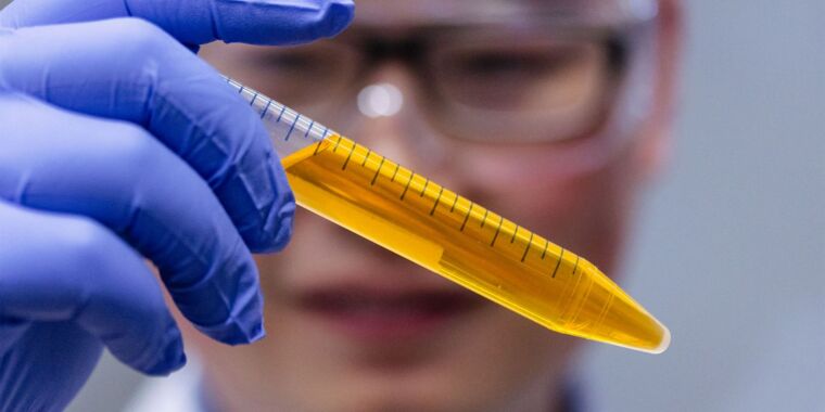
The developers of the system call each of these potentially modifiable spots on the template an epi-bit, with the modified version corresponding to a 1 in a conventional computer bit and the unmodified version corresponding to a 0. Because no synthesis is required, multiple bits can be written simultaneously. To read the information, the scientists rigged the system so that 1s fluoresce and 0s don’t. The fluorescence, along with the sequences of bases, was read as the DNA was passed through a tiny pore.
Pictures in a meta-genome
Using this system, Zhang et al. created five DNA templates and 175 bricks to record 350 bits at a time. Using a collection of tagged template molecules, the researchers could store and read roughly 275,000 bits, including a color picture of a panda’s face and a rubbing of a tiger from the Han dynasty, which ruled China from 202 BCE to 220 CE.
They then had 60 student volunteers “with diverse academic backgrounds” store texts of their choice in epi-bits using a simple kit in a classroom. Twelve of the 15 stored texts were read successfully.
We’re not quite ready for your cat videos yet, though. There are still errors in the printing and reading steps, and since these modifications don’t survive when DNA is copied, making additional versions of the stored information may get complicated. Plus, the stability of these modifications under different storage conditions remains unknown, although the authors note that their epi-bits stayed stable at temperatures of up to 95o° C.
But once these and a few other problems are solved—and the technology is scaled up, further optimized and automated, and/or tweaked to accommodate other types of epigenetic modifications—it will be a clever and novel way to harness natural data storage methods for our needs.
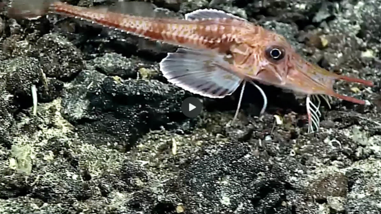
Finding out what controls the formation of sensory legs meant growing sea robins from eggs. The research team observed that the legs of sea robins develop from the three pectoral fin rays that are around the stomach area of the fish, then separate from the fin as they continue to develop. Among the most active genes in the developing legs is the transcription factor (a protein that binds to DNA and turns genes on and off) known as tbx3a. When genetically engineered sea robins had tbx3a edited out with CRISPR-Cas9, it resulted in fewer legs, deformed legs, or both.
“Disruption of tbx3a results in upregulation of pectoral fin markers prior to leg separation, indicating that leg rays become more similar to fins in the absence of tbx3a,” the researchers said in a second study, also published in Current Biology.
To see whether genes for sensory legs are a dominant feature, the research team also tried creating sea robin hybrids, crossing species with and without sensory legs. This resulted in offspring with legs that had sensory capabilities, indicating that it’s a genetically dominant trait.
Exactly why sea robins evolved the way they did is still unknown, but the research team came up with a hypothesis. They think the legs of sea robin ancestors were originally intended for locomotion, but they gradually started gaining some sensory utility, allowing the animal to search the visible surface of the seafloor for food. Those fish that needed to search deeper for food developed sensory legs that allowed them to taste and dig for hidden prey.
“Future work will leverage the remarkable biodiversity of sea robins to understand the genetic basis of novel trait formation and diversification in vertebrates,” the team also said in the first study. “Our work represents a basis for understanding how novel traits evolve.”
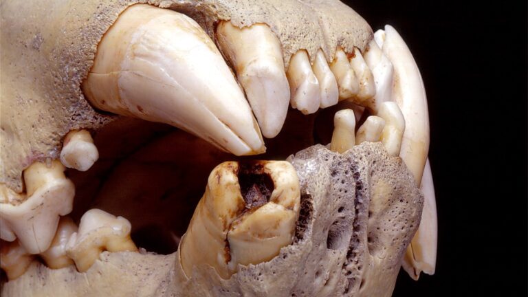
For several months in 1898, a pair of male lions turned the Tsavo region of Kenya into their own human hunting grounds, killing many construction workers who were building the Kenya-Uganda railway. A team of scientists has now identified exactly what kinds of prey the so-called “Tsavo Man-Eaters” fed upon, based on DNA analysis of hairs collected from the lions’ teeth, according to a recent paper published in the journal Current Biology. They found evidence of various species the lions had consumed, including humans.
The British began construction of a railway bridge over the Tsavo River in March 1898, with Lieutenant-Colonel John Henry Patterson leading the project. But mere days after Patterson arrived on site, workers started disappearing or being killed. The culprits: two maneless male lions, so emboldened that they often dragged workers from their tents at night to eat them. At their peak, they were killing workers almost daily—including an attack on the district officer, who narrowly escaped with claw lacerations on his back. (His assistant, however, was killed.)
Patterson finally managed to shoot and kill one of the lions on December 9 and the second 20 days later. The lion pelts decorated Patterson’s home as rugs for 25 years before being sold to Chicago’s Field Museum of Natural History in 1924. The skins were restored and used to reconstruct the lions, which are now on permanent display at the museum, along with their skulls.
Tale of the teeth
The Tsavo Man-Eaters naturally fascinated scientists, although the exact number of people they killed and/or consumed remains a matter of debate. Estimates run anywhere from 28–31 victims to 100 or more, with a 2009 study that analyzed isotopic signatures of the lions’ bone collagen and hair keratin favoring the lower range.
Over the last few years, Virginia Tech scientists have been looking to the octopus for inspiration to design technologies that can better grip a wide variety of objects in underwater environments. Their latest breakthrough is a special switchable adhesive modeled after the shape of the animal’s suckers, according to a new paper published in the journal Advanced Science.
“I am fascinated with how an octopus in one moment can hold something strongly, then release it instantly. It does this underwater, on objects that are rough, curved, and irregular—that is quite a feat,” said co-author and research group leader Michael Bartlett. “We’re now closer than ever to replicating the incredible ability of an octopus to grip and manipulate objects with precision, opening up new possibilities for exploration and manipulation of wet or underwater environments.”
As previously reported, there are several examples in nature of efficient ways to latch onto objects in underwater environments, per the authors. Mussels, for instance, secrete adhesive proteins to attach themselves to wet surfaces, while frogs have uniquely structured toe pads that create capillary and hydrodynamic forces for adhesion. But cephalopods like the octopus have an added advantage: The adhesion supplied by their grippers can be quickly and easily reversed, so the creatures can adapt to changing conditions, attaching to wet and dry surfaces.
From a mechanical engineering standpoint, the octopus has an active, pressure-driven system for adhesion. The sucker’s wide outer rim creates a seal with the object via a pressure differential between the chamber and the surrounding medium. Then muscles (serving as actuators) contract and relax the cupped area behind the rim to add or release pressure as needed.
There have been several attempts to mimic cephalopods when designing soft robotic grippers, for example. Back in 2022, Bartlett and his colleagues wanted to go one step further and recreate not just the switchable adhesion but also the integrated sensing and control. The result was Octa-Glove, a wearable system for gripping underwater objects that mimicked the arm of an octopus.
Improving the Octa-Glove
Grabbing and releasing underwater objects of different sizes and shapes with an octopus-inspired adhesive. Credit: Chanhong Lee and Michael Bartlett
For the adhesion, they designed silicone stalks capped with a pneumatically controlled membrane, mimicking the structure of octopus suckers. These adhesive elements were then integrated with an array of LIDAR optical proximity sensors and a micro-control for the real-time detection of objects. When the sensors detect an object, the adhesion turns on, mimicking the octopus’s nervous and muscular systems. The team used a neoprene wetsuit glove as a base for the wearable glove, incorporating the adhesive elements and sensors in each finger, with flexible pneumatic tubes inserted at the base of the adhesive elements.
Based on the stereotypical hairpin structure, researchers have scanned genomes and found over 38,000 likely precursors; nearly 50,000 mature microRNAs have been discovered by sequencing all the RNA found in cells from a variety of species. While found widely in animals, they’ve also been discovered in plants, raising the possibility that they existed in a single-celled ancestral organism.
While some microRNA genes, including lin-4 and let-7, have dramatic phenotypes when mutated, many have weak or confusing effects. This is likely in part due to the fact that a single microRNA can bind to and regulate a variety of genes and so may have a mix of effects when mutated. In other cases, several different microRNAs may bind to the same messenger RNA, creating a redundancy that makes the loss of a single microRNA difficult to detect.
Nevertheless, there’s plenty of evidence that, collectively, they’re essential for normal development in many organisms and tissues. Knocking out the gene that encodes the Dicer protein, which is needed for forming mature microRNAs, causes early embryonic lethality. Knockouts of the gene in specific cell types cause a variety of defects. For example, B cells never mature if Dicer is lost in that cell lineage, and a knockout in nerve cells causes microcephaly and limiting branching of connections among neurons, leading the animals to die shortly after birth.
This being the Medicine prize, the Nobel Committee also cite a number of human genetic diseases that are caused by mutations in microRNA genes.
Overall, the award highlights just how complex life is at the cellular level. There’s a fair number of genes that have to be made by every cell simply to enable their survival. But as for the rest, they exist embedded in complex regulatory networks that interact to ensure that proteins are made only where and when they’re needed, and often degraded if they somehow get made anyway. And every now and then, fundamental research in an oddball species is still telling us unexpected things about those networks.
In 2014, researchers monitoring acoustic recordings from the Mariana Archipelago picked up an unusual whale vocalization with both low- and high-frequency components. It seemed to be a whale call, but it sounded more mechanical than biological and has since been dubbed a “biotwang.”
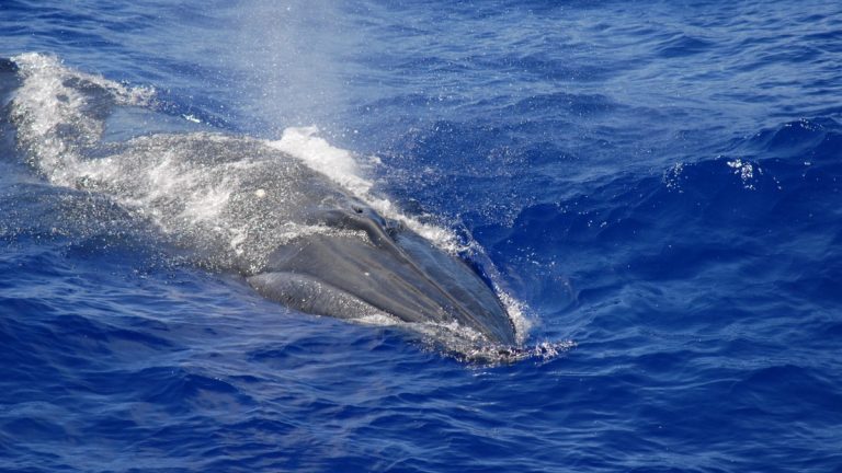
Now a separate team of scientists has developed a machine-learning model to scan a dataset of recordings of whale vocalizations from various species to help identify the source of such calls. Combining that analysis with visual observations allowed the team to identify the source of the biotwang: a species of baleen whales called Bryde’s (pronounced “broodus”) whales. This should help researchers track populations of these whales as they migrate to different parts of the world, according to a recent paper published in the journal Frontiers in Marine Science.
Marine biologists often rely on a powerful tool called passive acoustic monitoring for long-term data collection of the ocean’s acoustic environment, including whale vocalizations. Bryde’s whale calls tend to be regionally specific, per the authors. For instance, calls in the eastern North Pacific are pretty well documented, with frequencies typically falling below 100 Hz, augmented by harmonic frequencies as high as 400 Hz. Far less is known about the sounds made by Bryde’s whales in the western and central North Pacific, since for many years there were only three known recordings of those vocalizations—including a call dubbed “Be8” (starting at 45 Hz with multiple harmonics) and mother-calf calls.
That changed with the detection of the biotwang in 2014. It’s quite a distinctive, complex call that typically lasts about 3.5 seconds, with five stages, starting at around 30 Hz and ending with a metallic sound that can reach as high as 8,000 Hz. “It’s a real weird call,” co-author Ann Allen, a scientist at NOAA Fisheries, told Ars. “Anybody who wasn’t familiar with whales would think it was some sort of artificial sound, made by a naval ship.” The 2014 team was familiar with whale vocalizations and originally attributed the strange sound to baleen whales. But that particular survey was autonomous, and without accompanying visual observations, the scientists could not definitively confirm their hypothesis.
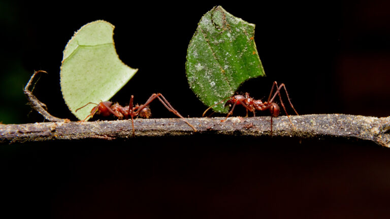
Tracing the lineages of agricultural ants to their most recent common ancestor revealed that the ancestor probably lived through the end-Cretaceous mass extinction—the one that killed off the dinosaurs. The researchers argue that the two were almost certainly related. Current models suggest that there was so much dust in the atmosphere after the impact that set off the mass extinction that photosynthesis shut down for nearly two years, meaning minimal plant life. By contrast, the huge amount of dead material would allow fungi to flourish. So, it’s not surprising that ants started to adapt to use what was available to them.
That explains the huge cluster of species that cooperate with fungi. However, most of the species that engage in organized farming don’t appear until roughly 35 million years after the mass extinction, at the end of the Eocene (that’s about 33 million years before the present period). The researchers suggest that the climate changes that accompanied the transition to the Oligocene included a drying out of the tropical Americas, where the fungus-farming ants had evolved. This would cut down on the availability of fungi in the wild, potentially selecting for the ability of species that could propagate fungal species on their own.
This also corresponds to the origins of the yeast strains used by farming ants, as well as the most specialized agricultural fungal species. But it doesn’t account for the origin of coral fungus farmers, which seems to have occurred roughly 10 million years later.
The work gives us a much clearer picture of the origin of agriculture in ants and some reasonable hypotheses regarding the selective pressures that might have led to its evolution. In the long term, however, the biggest advance here may be the resources generated during this study. Ultimately, we’d like to understand the genetic basis for the changes in the ants’ behavior, as well as how the fungi have adapted to better provide for their farmers. To do that, we’ll need to compare the genomes of agricultural species with their free-living relatives. The DNA gathered for this study will ultimately be needed to pursue those questions.
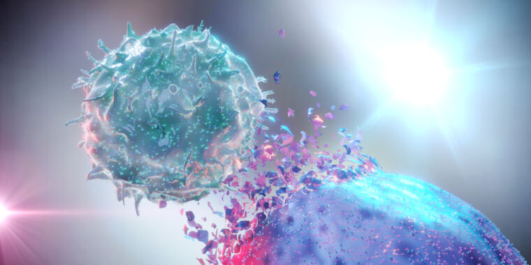
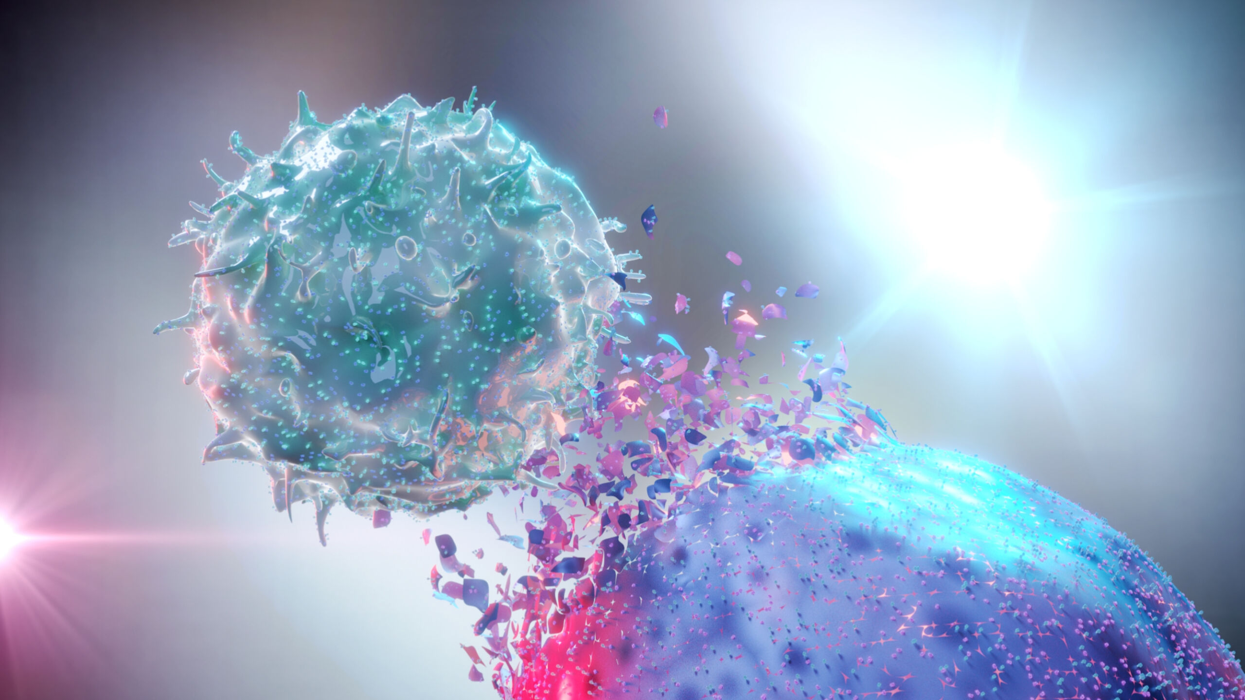
Enlarge/ 3D rendering of an NK Cell destroying a cancer cell.
Billions of cells die in your body every day. Some go out with a bang, others with a whimper.
They can die by accident if they’re injured or infected. Alternatively, should they outlive their natural lifespan or start to fail, they can carefully arrange for a desirable demise, with their remains neatly tidied away.
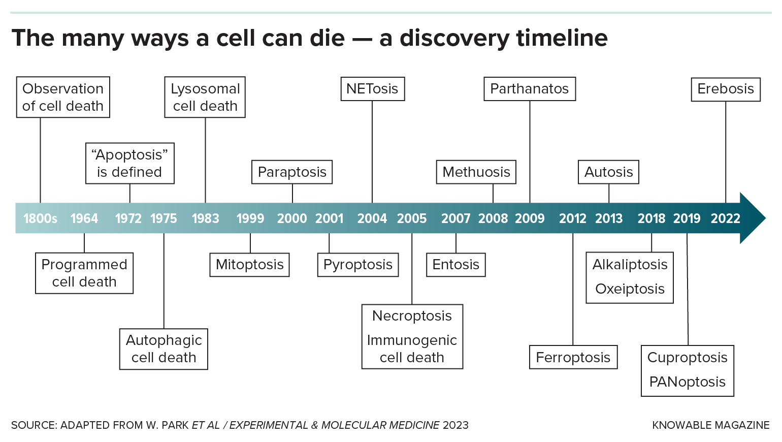
Originally, scientists thought those were the only two ways an animal cell could die, by accident or by that neat-and-tidy version. But over the past couple of decades, researchers have racked up many more novel cellular death scenarios, some specific to certain cell types or situations. Understanding this panoply of death modes could help scientists save good cells and kill bad ones, leading to treatments for infections, autoimmune diseases, and cancer.
“There’s lots and lots of different flavors here,” says Michael Overholtzer, a cell biologist at Memorial Sloan Kettering Cancer Center in New York. He estimates that there are now more than 20 different names to describe cell death varieties.
Here, Knowable Magazine profiles a handful of classic and new modes by which cells kick the bucket.
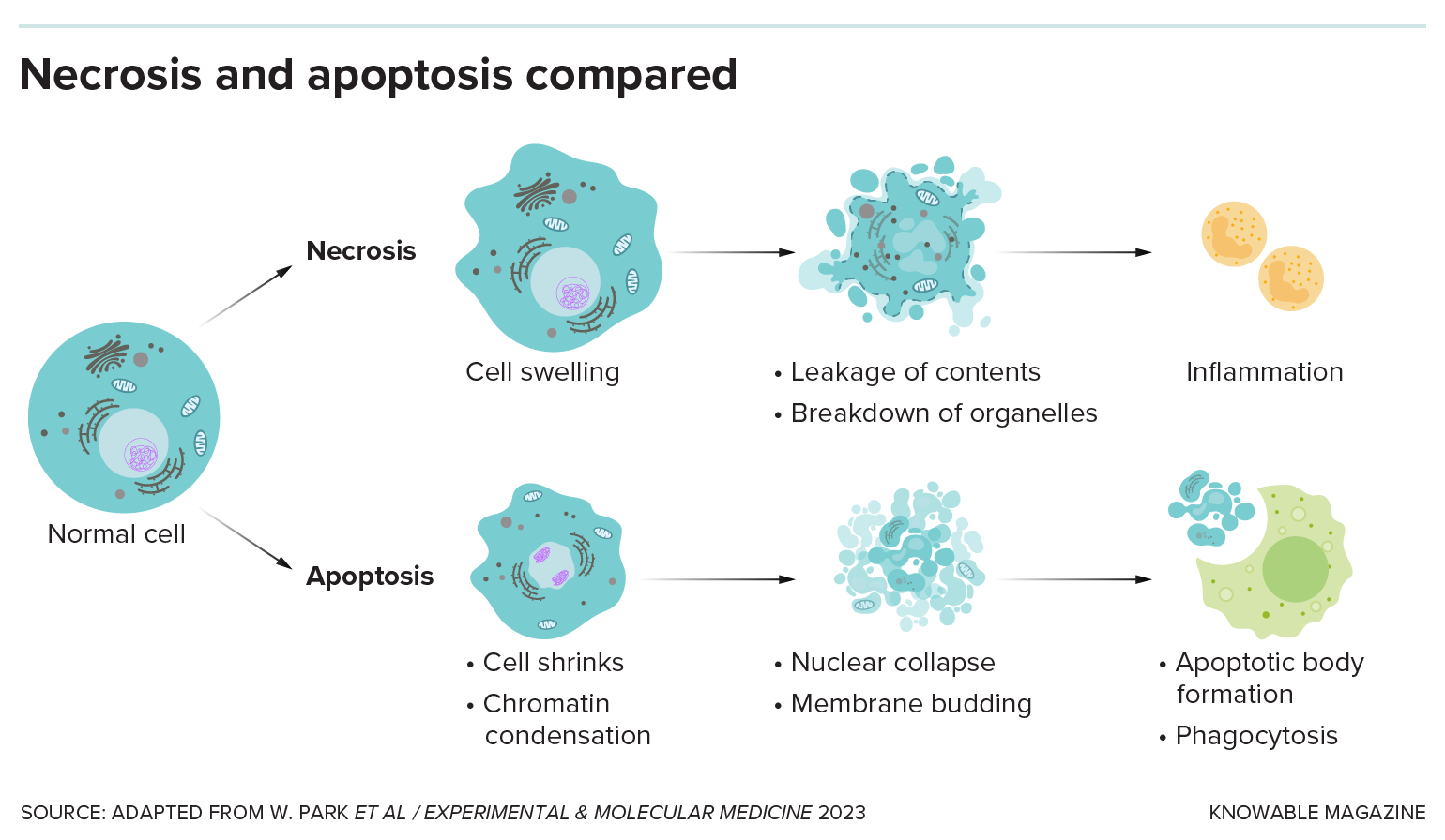
Unplanned cell death: Necrosis
Lots of bad things can happen to cells: They get injured or burned, poisoned or starved of oxygen, infected by microbes or otherwise diseased. When a cell dies by accident, it’s called necrosis.
There are several necrosis types, none of them pretty: In the case of gangrene, when cells are starved for blood, cells rot away. In other instances, dying cells liquefy, sometimes turning into yellow goop. Lung cells damaged by tuberculosis turn smushy and white — the technical name for this type, “caseous” necrosis, literally means “cheese-like.”
Any form of death other than necrosis is considered “programmed,” meaning it’s carried out intentionally by the cell because it’s damaged or has outlived its usefulness.
A good, clean death: Apoptosis
The two main categories of programmed cell death are “silent and violent,” says Thirumala-Devi Kanneganti, an immunologist at St. Jude Children’s Research Hospital in Memphis, Tennessee. Apoptosis, first named in 1972, is the original silent type: It’s a neat, clean form of cell death that doesn’t wake the immune system.
That’s handy when cells are damaged or have served out their purpose. Apoptosis allows tadpoles to discard tail cells when they become frogs, for example, or human embryos to dispose of the webbing between developing fingers.
The cell shrinks and detaches from its neighbors. Genetic material in the nucleus breaks into pieces that scrunch together, and the nucleus itself fragments. The membrane bubbles and blisters, and the cell disintegrates. Other cells gobble up the bits, keeping the tissue tidy.
Enlarge/ In necrosis, a cell dies by accident, releasing its contents and drawing immune cells to the site of damage by creating inflammation. In apoptosis, the cell collapses in on itself and the bits are cleared away without causing damaging inflammation.
Enlarge/ “When you spend more time putting electrodes back on than you do actually recording the EEGs, you get creative.”
Alienor Delsart
Our feline overlords aren’t particularly known for obeying commands from mere humans, which can make it difficult to study their behaviors in controlled laboratory settings. So a certain degree of ingenuity is required to get usable results—like crocheting adorable little hats for kitties taking part in electroencephalogram (EEG) experiments. That’s what researchers at the University of Montreal in Quebec, Canada, did to learn more about assessing chronic pain in cats—and they succeeded. According to their recent paper published in the Journal of Neuroscience Methods, it’s the first time scientists have recorded the electrical activity in the brains of conscious cats.
According to the authors, one-quarter of adult cats suffer from osteoarthritis and chronic pain that worsens with age. There are currently limited treatment options, namely, non-steroidal anti-inflammatory drugs, which can have significant side effects for the cats. An injectable monoclonal antibody tailored for cats has recently been developed to neutralize excessive nerve growth factor, but other alternative treatment options like supplements and regenerative medicine have yet to be tested. Nor has the effectiveness of certain smells or lighting in altering pain perception in felines been tested.
That was the Montreal team’s primary objective for their experiments. Initially, they tried to place electrodes on the heads of 11 awake adult cats with osteoarthritis, but the cats kept shaking off the electrodes.
“When you spend more time putting electrodes back on than you do actually recording the EEGs, you get creative,” co-author Aliénor Delsart of the University of Montreal told New Scientist. So he and his co-authors tapped a graduate student with crocheting skills to make the little hats. Not only did the hats hold the electrodes in place, but the cats also stopped trying to chew the wires.
With that problem solved, the real experiments could begin, designed to record brain activity of cats in response to smelling certain substances or seeing different wavelengths of colored light. The kitty subjects were housed as a group in an environment with lighting, temperature, and humidity controls, along with perches, beds, scratching posts, and cat toys.
Electrodes were attached with no need to shave the cats’ hair, thanks to a conductive paste to improve electrode/skin contact. First they recorded the basal activity before moving to exposure to sensory stimuli: a grapefruit smell for olfactory stimulation, and red, blue, and green lighting in a darkened room for visual stimulation.
Granted, there were still a few motion artifacts in that data; two cats were excluded from the data analysis for that reason. And the authors acknowledged the small sample size and largely descriptive nature of their analysis, which they deemed appropriate for what is essentially a test of the feasibility of their approach. The study met the group’s primary objectives: to assess whether the EEG method was feasible with conscious cats and whether the resulting analytical methods were an efficient means to characterize how the cats responded to specific sensory stimuli. “This opens new avenues for investigating chronic pain mechanisms and developing novel therapeutic strategies,” the authors concluded.
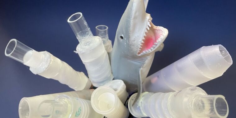
Enlarge/ Shark intestines are naturally occurring Tesla valves; scientists have figured out how to mimic their unique structure.
Sarah L. Keller/University of Washington
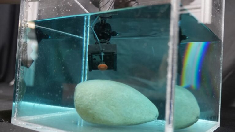
Scientists at the University of Washington have re-created the distinctive spiral shapes of shark intestines in 3D-printed pipes in order to study the unique fluid flow inside the spirals. Their prototypes kept fluids flowing in one preferred direction with no need for flaps to control that flow and performed significantly better than so-called “Tesla valves,” particularly when made of soft polymers, according to a new paper published in the Proceedings of the National Academy of Sciences.
As we’ve reported previously, in 1920, Serbian-born inventor Nikola Tesla designed and patented what he called a “valvular conduit“: a pipe whose internal design ensures that fluid will flow in one preferred direction, with no need for moving parts, making it ideal for microfluidics applications, among other uses. The key to Tesla’s ingenious valve design is a set of interconnected, asymmetric, tear-shaped loops.
In his patent application, Tesla described this series of 11 flow-control segments as being made of “enlargements, recessions, projections, baffles, or buckets which, while offering virtually no resistance to the passage of fluid in one direction, other than surface friction, constitute an almost impassable barrier to its flow in the opposite direction.” And because it achieves this with no moving parts, a Tesla valve is much more resistant to the wear and tear of frequent operation.
Tesla claimed that water would flow through his valve 200 times slower in one direction than another, which may have been an exaggeration. A team of scientists at New York University built a working Tesla valve in 2021, in accordance with the inventor’s design, and tested that claim by measuring the flow of water through the valve in both directions at various pressures. The scientists found the water only flowed about two times slower in the nonpreferred direction.
Flow rate proved to be a critical factor. The valve offered very little resistance at slow flow rates, but once that rate increased above a certain threshold, the valve’s resistance would increase as well, generating turbulent flows in the reverse direction, thereby “plugging” the pipe with vortices and disruptive currents. So it actually works more like a switch and can also help smooth out pulsing flows, akin to how AC/DC converters turn alternating currents into direct currents. That may even have been Tesla’s original intent in designing the valve, given that his biggest claim to fame is inventing both the AC motor and an AC/DC converter.
It helps to be a shark
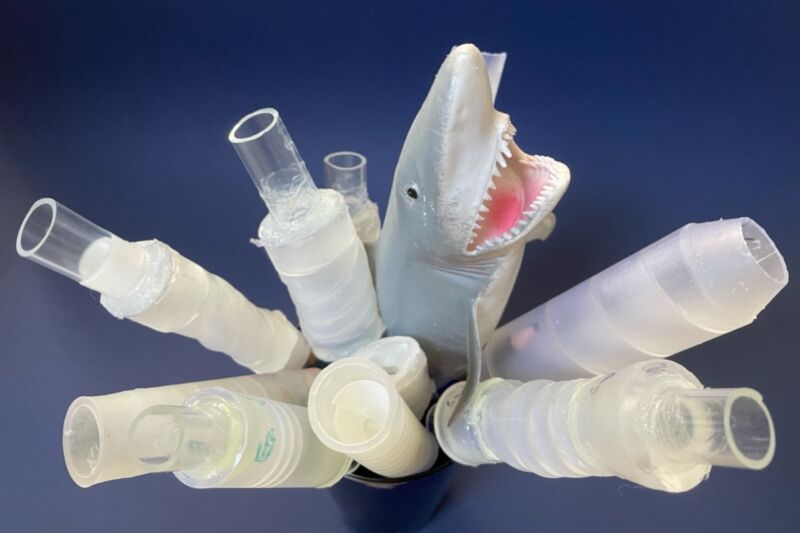
Enlarge/ Different kinds of sharks have intestines with different spiral patterns that favor fluid flow in one direction.
Ido Levin
The Tesla valve also provides a useful model for how food moves through the digestive system of many species of shark. In 2020, Japanese researchers reconstructed micrographs of histological sections from a species of catshark into a three-dimensional model, offering a tantalizing glimpse of the anatomy of a scroll-type spiral intestine. The following year, scientists took CT scans of shark intestines and concluded that the intestines are naturally occurring Tesla valves.
That’s where the work of UW postdoc Ido Levin and his co-authors comes in. They had questions about the 2021 research in particular. “Flow asymmetry in a pipe with no moving flaps has tremendous technological potential, but the mechanism was puzzling,” said Levin. “It was not clear which parts of the shark’s intestinal structure contributed to the asymmetry and which served only to increase the surface area for nutrient uptake.”
Levin et al. 3D-printed several pipes with an internal helical structure mimicking that of shark intestines, varying certain geometrical parameters like the number of turns or the pitch angle of the helix. It was admittedly an idealized structure, so the team was delighted when the first batch, made from rigid materials, produced the hoped-for flow asymmetry. After further fine-tuning of the parameters, the rigid printed pipes produced flow asymmetries that matched or exceeded Tesla valves.
Enlarge/ Eight of the team’s 3D-printed prototypes with various interior helices.
Ido Levin/University of Washington
But the researchers weren’t done yet. “[Prior work] showed that if you connect these intestines in the same direction as a digestive tract, you get a faster flow of fluid than if you connect them the other way around. We thought this was very interesting from a physics perspective,” said Levin last year while presenting preliminary results at the 67th Annual Biophysical Society Meeting. “One of the theorems in physics actually states that if you take a pipe, and you flow fluid very slowly through it, you have the same flow if you invert it. So we were very surprised to see experiments that contradict the theory. But then you remember that the intestines are not made out of steel—they’re made of something soft, so while fluid flows through the pipe, it deforms it.”
That gave Levin et al. the idea to try making their pipes out of soft deformable polymers—the softest commercially available ones that could also be used for 3D printing. That batch of pipes performed seven times better on flow asymmetry than any prior measurements of Tesla valves. And since actual shark intestines are about 100 times softer than the polymers they used, the team thinks they can achieve even better performance, perhaps with hydrogels when they become more widely available as 3D printing continues to evolve. The biggest challenge, per the authors, is finding soft materials that can withstand high deformations.
Finally, because the pipes are three-dimensional, they can accommodate larger fluid volumes, opening up applications in larger commercial devices. “Chemists were already motivated to develop polymers that are simultaneously soft, strong and printable,” said co-author Alshakim Nelson, whose expertise lies in developing new types of polymers. “The potential use of these polymers to control flow in applications ranging from engineering to medicine strengthens that motivation.”
Enlarge/ Zihao Ou, who helped develop this solution, holds a tube of it.
One key challenge in medical imaging is to look past skin and other tissue that are opaque to see internal organs and structures. This is the reason we need things like ultrasonography, magnetic resonance, or X-rays. There are chemical clearing agents that can make tissue transparent, like acrylamide or tetrahydrofuran, but they are almost never used in living organisms because they’re either highly toxic or can dissolve away essential biomolecules.
But now, a team of Stanford University scientists has finally found an agent that can reversibly make skin transparent without damaging it. This agent was tartrazine, a popular yellow-orange food dye called FD&C Yellow 5 that is notably used for coloring Doritos.
Playing with light
We can’t see through the skin because it is a complex tissue comprising aqueous-based components such as cell interiors and other fluids, as well as protein and lipids. The refractive index is a value that indicates how much light slows down (on average, of course) while going through a material compared to going through a vacuum. The refractive index of those aqueous components is low, while the refractive index of the proteins and lipids is high. As a result, light traveling through skin constantly bends as it endlessly crosses the boundary between high and low refractive index materials.
This scatters the light—once it penetrates the skin, it never gets back. What we see is just the light that bounces off the skin’s surface. The trick to making things transparent is mostly about making their refractive index uniform, so light, or at least some part of the spectrum, doesn’t bend all the time and doesn’t get scattered. This is exactly where the Doritos dye came in.
“The most surprising part of this study is that we usually expect dye molecules to make things less transparent,” says Guosong Hong, an assistant professor of materials science and engineering at Stanford and senior author of the paper. “For example, if you mix blue pen ink in water, the more ink you add, the less light can pass through the water. However, in our experiment, when we dissolve tartrazine in an opaque material like muscle or skin, which normally scatters light, the more tartrazine we add, the clearer the material becomes. This goes against what we typically expect with dyes.”
Transparency lotion
Hong’s team simply dissolved the dye in an aqueous solution and created a transparency-inducing lotion of sorts. It worked, because the dye reduced the difference in refractive index between water and lipids in the skin. Then the team started massaging it gently into a bit of polymer gel that emulated the light-scattering properties of tissue. From there, they moved to thinly sliced chicken breasts and to live mice.
The “transparency lotion” needed just a few minutes to start working when applied to a mouse’s skin. Massaged into a shaven scalp, it lets the scientists see the cerebral blood vessels with laser speckle contrast imaging, a technique that normally requires removal of the scalp to work. When applied to the mouse’s abdomen, it made all the internal organs, including the liver, bladder, and small intestine, visible to the naked eye. All that was needed to reverse the effect and make the skin opaque again was washing the lotion off with water.
There were some problems, though. One of them was that tartrazine absorbed most light at wavelengths around 257 and 428 nanometers, which let us see shades of violet and blue. On the other hand, it had minimal absorption above 600 nanometers, which meant that the transparent skin tinted everything red. The second issue was the depth of penetration. The lotion worked well only at spots where the skin was thin, and couldn’t penetrate deep enough where the skin was thicker.
Finally, its formulation was not universal. It relied on finding a chemical that could match the refractive index of lipids when dissolved in water, but the exact composition of the lotion was determined through trial and error. If there’s a lot of mouse-to-mouse variation, it might make it hard to come up with a one-size-fits-all solution.
Tattoos and needles
The problem of penetrating deeper into thick skin was partially solved by making the application a bit more painful. “Using microneedle patch applicators or subcutaneous injections could help deliver the molecules through thicker layers of skin,” Hong explains. The red tint issue, he suggested, might be handled by testing different dyes. “The research in my lab is currently focused on identifying molecules with sharp absorption in the near-ultraviolet region, minimizing spectral tailing into the visible range to ensure tissue transparency without the presence of a red tone,” Hong said.
“This study has only been conducted on animals. However, if the same technique could be applied to humans, it could offer a variety of benefits in biology, diagnostics, and even cosmetics,” Hong suggests. The benefits he is focusing on include evaluating deep-seated tumors without relying on biopsies, making blood tests less stressful by making locating the veins easier, and even things like improved laser tattoo removals by allowing the pigment beneath the skin to be targeted precisely.
But there is some bad news. Even though the FD&C Yellow 5 dye is widely available, replicating Hong’s results at home and making the transparency lotion on your own is not the brightest idea. “We strongly discourage attempting this on the human skin, as the toxicology of dye molecules in humans, particularly when applied topically, has not been fully evaluated,” Hong says.
And, in the end, it might not even work. “The human skin is significantly thicker than mouse skin, with the stratum corneum, the outermost layer of the epidermis, serving as a substantial barrier that prevents effective delivery of molecules into the dermis,” Hong explains
For many creatures, having a limb caught in a predator’s mouth is usually a death sentence. Not starfish, though—they can detach the limb and leave the predator something to chew on while they crawl away. But how can they pull this off?
Starfish and some other animals (including lizards and salamanders) are capable of autonomy (shedding a limb when attacked). The biology behind this phenomenon in starfish was largely unknown until now. An international team of researchers led by Maurice Elphick, professor of Animal Physiology and Neuroscience at Queen Mary University of London, have found that a neurohormone released by starfish is largely responsible for detaching limbs that end up in a predator’s jaws.
So how does this neurohormone (specifically a neuropeptide) let the starfish get away? When a starfish is under stress from a predatory attack, this hormone is secreted, stimulating a muscle at the base of the animal’s arm that allows the arm to break off.
The researchers confirmed this neuropeptide “acts as an autotomy-promoting factor in starfish and such it is the first neuropeptide to be identified as a regulator of autotomy in animals,” as they said in a study recently published in Current Biology.
Holding on
Elphick’s team studied how the neuropeptide known as ArSK/CCK1 facilitates autonomy in the European Starfish, Asterias rubens. ArSK/CCK1 is already known to inhibit feeding behavior in A. rubens by causing the stomach to contract, and muscle contraction plays a role in limb loss. The researchers found that its ability to trigger contractions goes beyond feeding.
Starfish underwent an experiment that simulated conditions where a predator’s jaw clamped down on one arm. Clamps were placed on one of three sections on a single arm, either on the end, middle, or at the site in the base where autotomy is known to occur, also known as the autotomy plane. The starfish were then suspended by these clamps above a glass bowl of seawater. During the first part of the experiment, the starfish were left to react naturally, but during the second part, they were injected with ArSK/CCK1.
Without the injection, autotomy was seen mostly in animals that had arms that were clamped closest to the autotomy plane. There was not nearly as much of a reaction from starfish when the arms were clamped in the middle or end.
In the second half of the experiment, the clamping used before was combined with an injection of ArSK/CCK1. For comparison, some were injected with the related neuropeptide ArSK/CCK2. A staggering 85 percent of ArSK/CCK1-injected animals that were clamped in the middle of the arm or closer to the autotomy plane exhibited autonomy, and some autotomized additional arms. This only happened in about 27 percent of those injected with ArSK/CCK2.
Letting go
While ArSK/CCK1 proved to be the most effective chemical trigger for autotomy, its activity in the autotomy plane depends on certain aspects of a starfish’s anatomy.
Like all echinoderms, starfish have endoskeletons built of tiny bones, or ossicles, linked by muscles and collagen fibers that allow the animals to change posture and move. Two exclusive features only found in the autotomy plane allow this structure to break. Under the skin of the autotomy plane, there is a region where bundles of collagen fibers are positioned far apart to make breakage easier. The second of these features is a band of muscle close to the region of collagen bundles. Known as the tourniquet muscle, this muscle is responsible for the constriction that allows an arm in danger to fall off.
Analyzing starfish arm tissue while it was undergoing autotomy gave the scientists a new perspective on this process. Right after a starfish has its arm seized by a predator, ArSK/CCK1 tells nerves in the tourniquet muscle to start constricting in the region right by the autonomy plane. While this is happening, the collagen in the body wall in that region softens and breaks, and so do the muscles and ligaments that hold together ossicles. It is now thought that ArSK/CCK1 is also involved in the softening of this tissue that prepares it for breakage.
After starfish autotomize a limb, that limb eventually regenerates. The same happens in other animals that can use autotomy to their advantage (such as lizards, which also grow their tails back). In the future, finding out why some animals have the ability to regenerate may tell us why we either never evolved it or some of our ancestors lost the ability. Elphick acknowledged that there might still be other unidentified factors working together with ArSK/CCK1, but further insight could someday give us a clearer picture of this process.
“Autotomy is a key adaptation for survival that has evolved in several animal taxa,” the research team said in the same study, “[and] the findings of this study provide a seminal insight into the neural mechanisms that control this remarkable biological process,”


