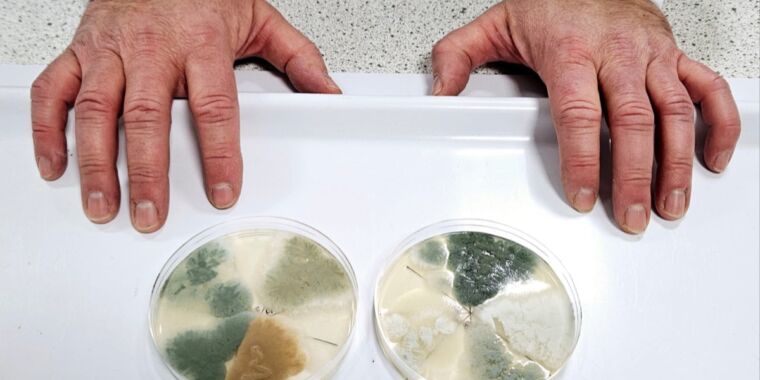
Exterminators keep getting calls for a reason. Wood-devouring insects, such as beetles, termites, and carpenter ants, are constantly chewing through walls or infecting trees and breaking them down. The fight against these insects usually involved noxious insecticides; but now, at least some of them can be eliminated using a certain species of fungus.
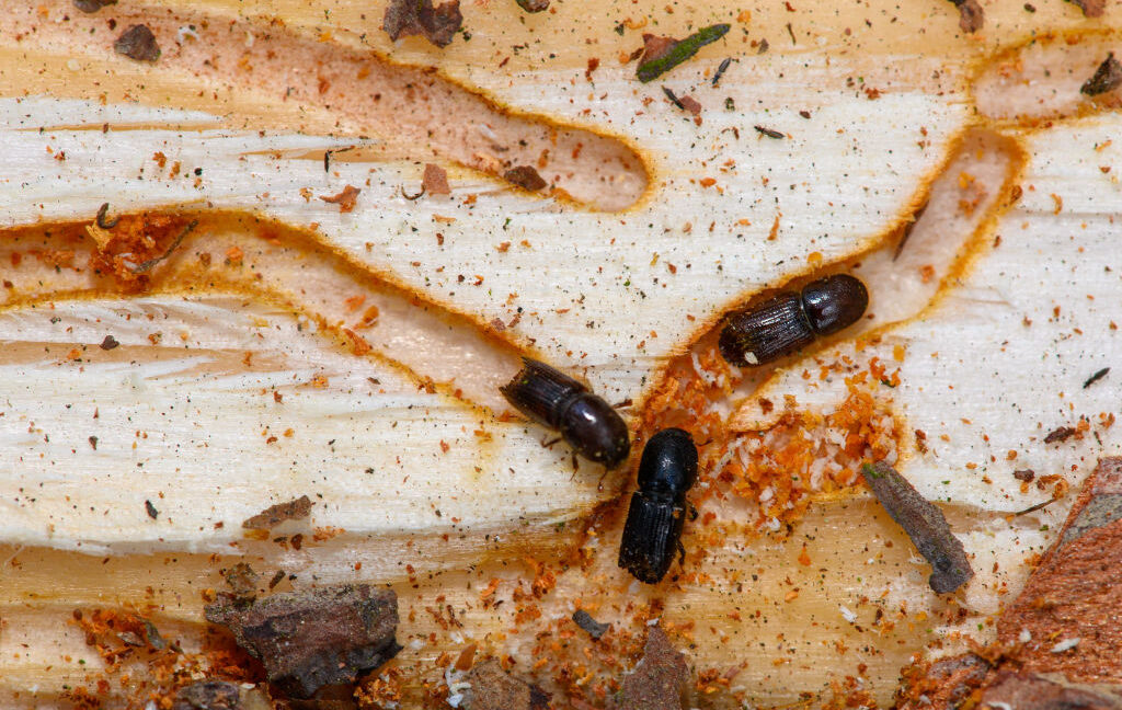
Infestations of bark beetles are the bane of spruce trees. Eurasian spruce bark beetles (Ips typographus) ingest bark high in phenolic compounds, organic molecules that often act as antioxidants and antimicrobials. They protect spruce bark from pathogenic fungi—and the beetles take advantage. Their bodies boost the antimicrobial power of these compounds by turning them into substances that are even more toxic to fungi. This would seem to make the beetles invulnerable to fungi.
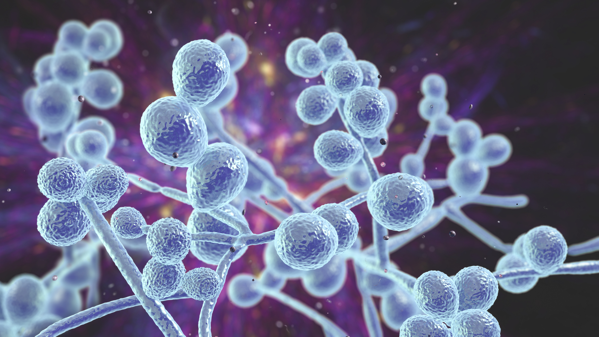
There is a way to get past the beetles’ borrowed defenses, though. Led by biochemist Ruo Sun, a team of researchers from the Max Planck Institute for Chemical Ecology in Jena, Germany, found that some strains of the fungus Beauveria bassiana are capable of infecting and killing the pests.
“Insect herbivores have long been known to accumulate plant defense metabolites from their diet as defenses against their own enemies,” she said in a study recently published in PNAS. “However, as shown here for B. bassiana, fungal pathogens are able to circumvent the toxicity of these dietary defenses and cause disease.”
First line of defense
Populations of bark beetles have recently exploded in temperate forests because of climate change. One species they feed on is the Norway spruce (Picea abies), which makes organic phenolic compounds known as stilbenes and flavonoids. Stilbenes are hydrocarbons that function as secondary metabolites for plants, and flavonoids, which are polyphenols, are also secondary plant metabolites that are often antioxidants. The spruce links both classes of compounds with sugars and relies on their antibacterial and antifungal activity.
When metabolized by the beetles, the spruce sugars are removed through hydrolysis, converting them into aglycones that are even more toxic to microscopic invaders. Despite that, some fungi appear to be able to deactivate these compounds. Strains of the fungal insect pathogen B. bassiana have been documented as killing some of these beetles in the wild.
Even with Fusarium graminearum, which has appeared on every continent but Antarctica, there is potential for introducing new genetic material into the environment that may exist in other countries but not the US and could have harmful consequences for crops.
How do you manage Fusarium graminearum infections?
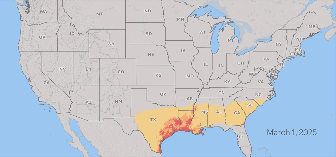
Fusarium graminearum infections generally occur during the plant’s flowering stage or when there is more frequent rainfall and periods of high humidity during early stages of grain production.
How Fusarium graminearum risk progressed in 2025. Yellow is low risk, orange is medium risk, and red is high risk. Fusarium Risk Tool/Penn State
Wheat in the southern US is vulnerable to infection during the spring. As the season advances, the risk from scab progresses north through the US and into Canada as the grain crops mature across the region, with continued periods of conducive weather throughout the summer.
Between seasons, Fusarium graminearum survives on barley, wheat, and corn plant residues that remain in the field after harvest. It reproduces by producing microscopic spores that can then travel long distances on wind currents, spreading the fungus across large geographic areas each season.
In wheat and barley, farmers can suppress the damage by spraying a fungicide onto developing wheat heads when they’re most susceptible to infection. Applying fungicide can reduce scab and its severity, improve grain weight, and reduce mycotoxin contamination.
However, integrated approaches to manage plant diseases are generally ideal, including planting barley or wheat varieties that are resistant to scab and also using a carefully timed fungicide application, rotating crops, and tilling the soil after harvest to reduce residue where Fusarium graminearum can survive the winter.
Even though fungicide applications may be beneficial, fungicides offer only some protection and can’t cure scab. If the environmental conditions are extremely conducive for scab, with ample moisture and humidity during flowering, the disease will still occur, albeit at reduced levels.
Fusarium Head Blight with NDSU’s Andrew Friskop.
Plant pathologists are making progress on early warning systems for farmers. A team from Kansas State University, Ohio State University, and Pennsylvania State University has been developing a computer model to predict the risk of scab. Their wheat disease predictive model uses historic and current environmental data from weather stations throughout the US, along with current conditions, to develop a forecast.
In areas that are most at risk, plant pathologists and commodity specialists encourage wheat growers to apply a fungicide during periods when the fungus is likely to grow to reduce the chances of damage to crops and the spread of mycotoxin.
Many insect species hear using tympanal organs, membranes roughly resembling our eardrums but located on their legs. Grasshoppers, mantises, and moths all have them, and for decades, we thought that female stinkbugs of the Dinidoridae family have them, too, although located a bit unusually on their hind rather than front legs.
Suspecting that they use their hind leg tympanal organs to listen to male courtship songs, a team of Japanese researchers took a closer look at the organs in Megymenum gracilicorne, a Dinidoridae stinkbug species native to Japan. They discovered that these “tympanal organs” were not what they seemed. They’re actually mobile fungal nurseries of a kind we’ve never seen before.
Portable gardens
Dinidoridae is a small stinkbug family that lives exclusively in Asia. The bug did attract some scientific attention, but not nearly as much as its larger relatives like Pentatomidae. Prior work looking specifically into organs growing on the hind legs of Dinidoridae females was thus somewhat limited. “Most research relied on taxonomic and morphological approaches. Some taxonomists did describe that female Dinidoridae stinkbugs have an enlarged part on the hind legs that looks like the tympanal organ you can find, for example, in crickets,” said Takema Fukatsu, an evolutionary biologist at the National Institute of Advanced Industrial Science and Technology in Tokyo.
Based on that appearance, these parts were classified as tympanal organs—the case was closed, and it stayed closed until Fukatsu’s team started examining them more closely. Most insects have tympanal organs on their front legs, not hind legs, or on abdominal segments. The initial goal of Fukatsu’s study was to figure out what impact this unusual position has on Dinidoridae females’ ability to hear sounds.
Early on in the study, it turned out that whatever Dinidoridae females have on their hind legs, they are not tympanal organs. “We found no tympanal membrane and no sensory neurons, so the enlarged parts on the hind legs had nothing to do with hearing,” Fukatsu explained. Instead, the organ had thousands of small pores filled with benign filamentous fungi. The pores were connected to secretory cells that released substances that Fukatsu’s team hypothesized were nutrients enabling the fungi to grow.
Fungi are an indispensable part of your microbiome, keeping the body’s host of microorganisms healthy as part of a system of checks and balances. But when you’re hit by an infection, fungi can be thrown out of equilibrium with other organisms inside you, leading to a more severe infection and other symptoms of illness.
For this reason, the pandemic immediately set off alarms for Iliyan Iliev, an immunologist at Weill Cornell Medical School. “We were thinking, the first thing that’s going to happen is people will start getting fungal co-infections,” he says. With the microbiome unbalanced, fungi might start running riot inside Covidpatients, Iliev reasoned. His fears were soon realized.
In research published in Nature Immunology, he and his team discovered that in patients with severe Covid, certain strains of gut fungi—knocked off-kilter by the virus—set off a prolonged immune response that could last long after the initial infection. This response potentially led to some of the respiratory symptoms experienced by these patients. These results, Iliev says, point to the critical role of the gut microbiome in the human immune response and could lead to better disease treatments down the line.
Imbalance of the gut microbiome has long been linked to disease. Ken Cadwell, an immunologist at the Perelman School of Medicine at the University of Pennsylvania, thinks of the microbiome as a metaphorical rainforest. “It’s a nice ecosystem—but if you cut down too many trees or bring in invasive species, you could make things go out of whack,” he says.
To see how the body’s internal fungi were affected during Covid and how this triggered the immune system, Iliev and his team started by looking at patients’ blood. After collecting samples from 91 people with Covid, they measured levels of antibodies against several fungi, to figure out if the body’s immune system was reacting against these. Significantly more anti-fungal antibodies, for instance, would indicate fungal overgrowth or invasion.
Takato Kusakabe, a postdoctoral fellow in Iliev’s lab and study author, ran plate after plate of experiments—a painstaking process—to quantify these antibody levels. The team found in patients with severe Covid, several fungi commonly found in the gut had increased antibodies against them (in comparison to uninfected people). Notably, these included Candida albicans, which is a common culprit of yeast infections. When the team then ran tests on fecal samples from 10 of the hospitalized Covid patients, these confirmed that the fungi being targeted by the antibodies were present in the patients’ guts—and at seemingly at higher levels than in uninfected controls, suggesting an imbalance in their microbiome.
Enlarge/ Scientists at the University of Nottingham have discovered how to create different colors of blue cheese.
University of Nottingham
Gourmands are well aware of the many varieties of blue cheese, known by the blue-green veins that ripple through the cheese. Different kinds of blue cheese have distinctive flavor profiles: they can be mild or strong, sweet or salty, for example. Soon we might be able to buy blue cheeses that belie the name and sport veins of different colors: perhaps yellow-green, reddish-brown-pink, or lighter/darker shades of blue, according to a recent paper published in the journal Science of Food.
“We’ve been interested in cheese fungi for over 10 years, and traditionally when you develop mould-ripened cheeses, you get blue cheeses such as Stilton, Roquefort, and Gorgonzola, which use fixed strains of fungi that are blue-green in color,” said co-author Paul Dyer of the University of Nottingham of this latest research. “We wanted to see if we could develop new strains with new flavors and appearances.”
Blue cheese has been around for a very long time. Legend has it that a young boy left his bread and ewe’s milk cheese in a nearby cave to pursue a lovely young lady he’d spotted in the distance. Months later, he came back to the cave and found it had molded into Roquefort. It’s a fanciful tale, but scholars think the basic idea is sound: people used to store cheeses in caves because their temperature and moisture levels were especially hospitable to harmless molds. That was bolstered by a 2021 analysis of paleofeces that found evidence that Iron Age salt miners in Hallstatt (Austria) between 800 and 400 BCE were already eating blue cheese and quaffing beer.
The manufacturing process for blue cheese is largely the same as for any cheese, with a few crucial additional steps. It requires cultivation of Penicillium roqueforti, a mold that thrives on exposure to oxygen. The P. roqueforti is added to the cheese, sometimes before curds form and sometimes mixed in with curds after they form. The cheese is then aged in a temperature-controlled environment. Lactic acid bacteria trigger the initial fermentation but eventually die off, and the P. roqueforti take over as secondary fermenters. Piercing the curds forms air tunnels in the cheese, and the mold grows along those surfaces to produce blue cheese’s signature veining.
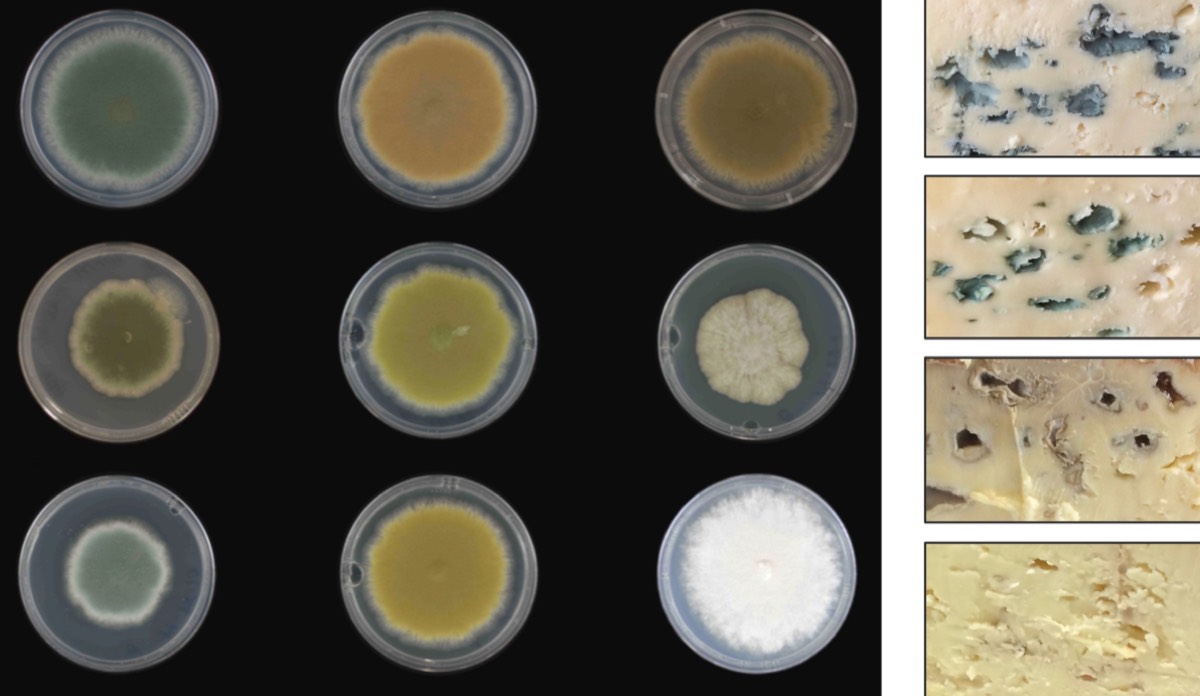
Once scientists published the complete genome for P. roqueforti, it opened up opportunities for studying this blue cheese fungus, per Dyer et al. Different strains “can have different colony cultures and textures, with commercial strains being sold partly on the basis of color development,” they wrote. This coloration comes from pigments in the coatings of the spores that form as the colony grows. Dyer and his co-authors set out to determine the genetic basis of this pigment formation in the hopes of producing altered strains with different spore coat colors.
The team identified a specific biochemical pathway, beginning with a white color that gradually goes from yellow-green, red-brown-pink, dark brown, light blue, and ultimately that iconic dark blue-green. They used targeted gene deletion to block pigment biosynthesis genes at various points in this pathway. This altered the spore color, providing a proof of principle without adversely affecting the production of flavor volatiles and levels of secondary metabolites called mycotoxins. (The latter are present in low enough concentrations in blue cheese so as not to be a health risk for humans, and the team wanted to ensure those concentrations remained low.)
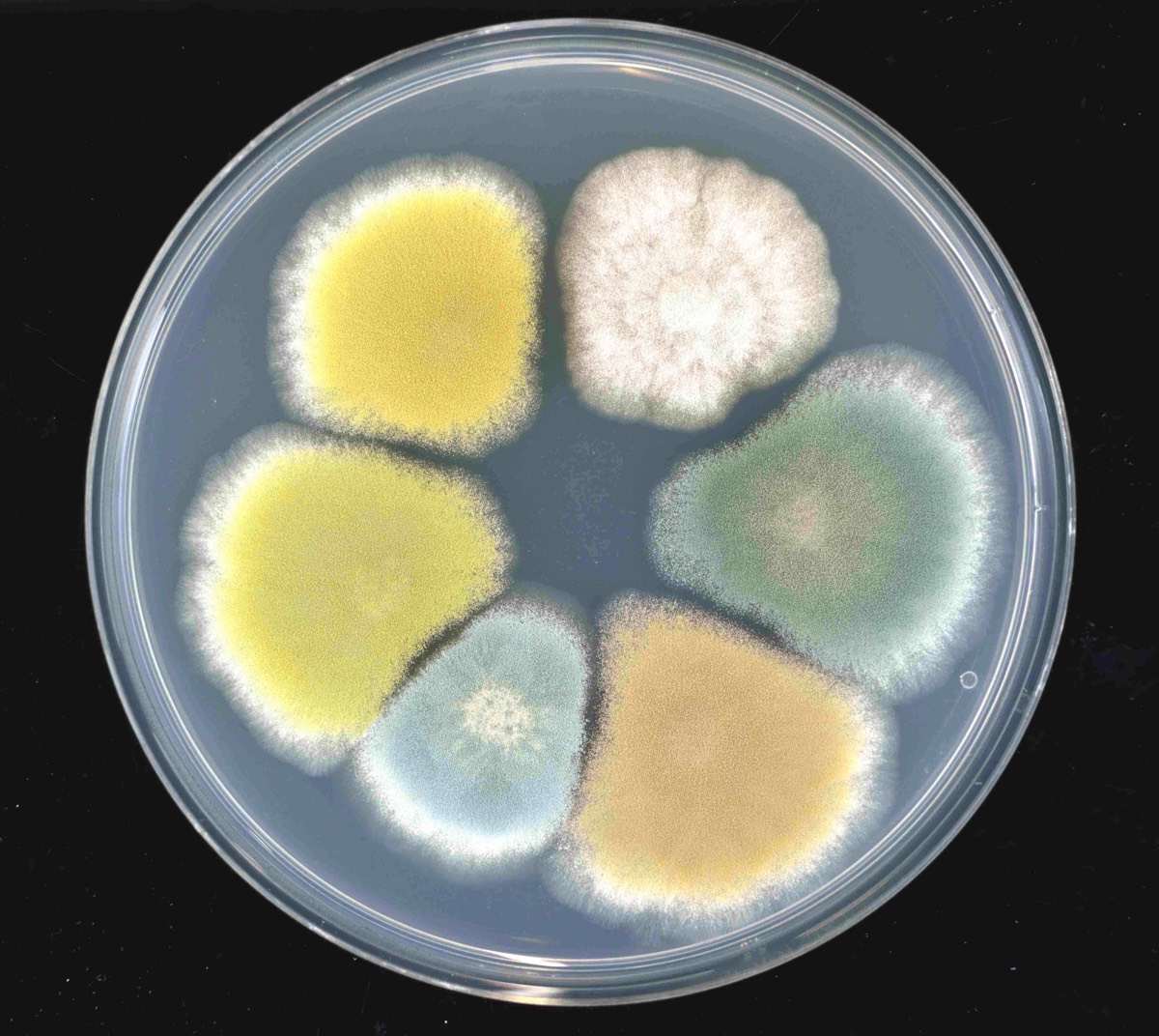
Enlarge/ (left) Spectrum of color strains produced in Pencillium roqueforti. (right) Cross sections of cheeses made with the original (dark blue-green) or new color (red-brown, bright green, white albino) strains of the fungus.
University of Nottingham
However, food industry regulations prohibit gene-deletion fungal strains for commercial cheese production. So Dyer et al. used UV mutagenesis—essentially “inducing sexual reproduction in the fungus,” per Dyer—to produce non-GMO mutant strains of the fungi to create “blue” cheeses of different colors, without increasing mycotoxin levels or impacting the volatile compounds responsible for flavor.
“The interesting part was that once we went on to make some cheese, we then did some taste trials with volunteers from across the wider university, and we found that when people were trying the lighter colored strains they thought they tasted more mild,” said Dyer. “Whereas they thought the darker strain had a more intense flavor. Similarly, with the more reddish-brown and a light green one, people thought they had a fruity, tangy element to them—whereas, according to the lab instruments, they were very similar in flavor. This shows that people do perceive taste not only from what they taste but also by what they see.”
Dyer’s team is hoping to work with local cheese makers in Nottingham and Scotland, setting up a spinoff company in hopes of commercializing the mutant strains. And there could be other modifications on the horizon. “Producers could almost dial up their list of desirable characteristics—more or less color, faster or slower growth rate, acidity differences,” Donald Glover of the University of Queensland in Australia, who was not involved in the research, told New Scientist.
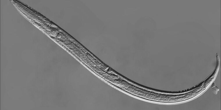
Some of the scariest monsters are microscopic. The carnivorous fungusArthrobotrys oligospora doesn’t seem like much while it’s eating away at rotting wood. But when it senses a live worm, it will trap its victim and consume it alive—pure nightmare fuel.
Until now, not much was known about how the attack of the killer fungus happens on a molecular level. Researchers from Academia Sinica in Taiwan have finally found out how the gene activity of the fungus changes when a nematode creeps too close to A. oligospora. Led by molecular biologist Hung-Che Lin, the research team discovered that the fungus synthesizes a sort of worm adhesive and additional trapping proteins to get ahold of its meal. It then produces enzymes that break down the worm so it can start feasting.
Caught in a trap
A.oligospora lives in the soil and is mostly saprotrophic, meaning it feeds on decaying organic matter. But that can quickly change if it finds itself deprived of nutrients or senses a tempting nematode nearby. This is when it goes into carnivore mode.
Lin and his colleagues wanted to see what happened when the fungus, low on nutrients, was introduced to the nematode Caenorhabiditis elegans. The fungus showed a significant increase in DNA replication when it sensed the worm. This resulted in trap cells having additional copies of the genome. The trap cells reside in fungal filaments, or hyphae, and produce a specialized worm adhesive that would allow those hyphae to stick to the worm once it was caught in the trap.
What may be the most important genetic actions in helping the fungus to create a trap out of hyphae is ribosome biogenesis, which enables increased protein production. Ribosomes are where proteins are made, so their biogenesis (literally the creation of more ribosomes) controls cell growth and also determines how much protein is synthesized.
The researchers also identified a new group of proteins, now known as Trap Enriched Proteins (TEPs), which were the most commonly produced proteins in fungal trap cells. These seemed to contribute to trap function rather than formation.
“Given TEP protein localization to the surface of trap cells, we hypothesized that TEPs may be critical for the function of the traps,” they said in a study recently published in PLoS Biology. “Adding C. elegans… leads to their immediate capture.”
As the fungus put more effort into creating a trap and forming worm adhesive, it deprioritized activities that are not really involved in the process. Segments of DNA that usually help A. oligospora digest dead matter were down-regulated, meaning there was lower gene activity on these segments in response to the fungus sensing the worm. When a worm came close to A. oligospora, the fungus showed an up-regulation of genes that produce proteases, or enzymes that break down proteins.
Can’t get out
Additional other genes didn’t see changes in activity until the worm was already caught. Once C. elegans entered the trap that A. oligospora had set with a sticky net of hyphae, the team noticed an increase in the production of proteins that weaken prey. These proteins are able to manipulate the cells of their prey so those cells function differently, potentially providing a way for the pathogen to break in and take over. The fungus then uses proteases to digest nematodes that get stuck in its hyphae.
A. oligospora has over 400 genes that encode proteins that control its interactions with other organisms. When the introduction of a nematode made the fungus go carnivorous, more than half of these started to behave differently. These proteins weaken C. elegans through a variety of mechanisms. To give one example, some of them fight off antimicrobial peptides produced by the nematode.
The adhesive synthesized by the fungus, now thought to have a close association with TEP proteins, may have no effect on humans but is a superglue for worms that binds hyphae to their flesh. They have no way of worming their way out of being eaten alive.
This experiment might have been ghastly for the nematodes involved, but it was a breakthrough for Lin’s team. They have now identified an entire new group of genes that make a fungal trap function. Their findings with A. oligospora could be compared to the gene activity of other pathogenic fungi, including those that destroy crops, so an improved generation of antifungals might someday be influenced by this microscopic horror movie.


