From chickens to humans, animals think “bouba” sounds round
Does “bouba” sound round to you? How about “maluma”? Neither are real words, but we’ve known for decades that people who hear them tend to associate them with round objects. There have been plenty of ideas put forward about why that would be the case, and most of them have turned out to be wrong. Now, in perhaps the weirdest bit of evidence to date, researchers have found that even newly hatched chickens seem to associate “bouba” with round shapes.
The initial finding dates all the way back to 1947, when someone discovered that people associated some word-like sounds with rounded shapes, and others with spiky ones. In the years since, that association got formalized as the bouba/kiki effect, received a fair bit of experimental attention, and ended up with an extensive Wikipedia entry.
One of the initial ideas to explain it was similarity to actual words (either phonetically or via the characters used to spell them), but then studies with speakers of different languages and alphabets showed that it is likely a general human tendency. The association also showed up in infants as young as 4 months old, well before they master speaking or spelling. Attempts to find the bouba/kiki effects in other primates, however, came up empty. That led to some speculation that it might be evidence of a strictly human processing ability that underlies our capacity to learn sophisticated languages.
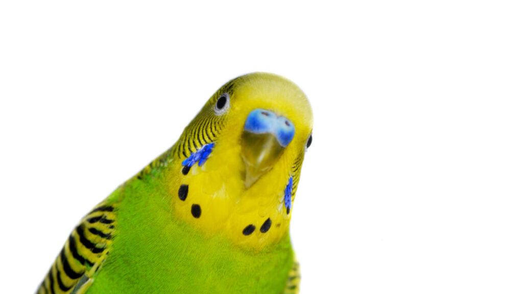
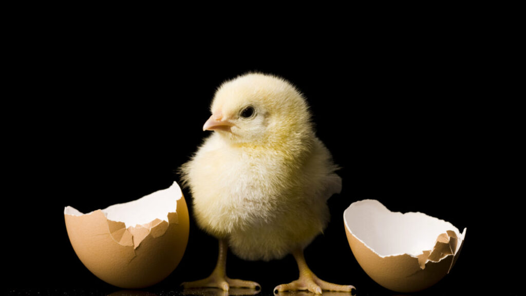
A team of Italian researchers—Maria Loconsole, Silvia Benavides-Varela, and Lucia Regolin—now have evidence that that isn’t true either. They decided to look for the bouba/kiki effect well beyond primates, instead turning to newly hatched chickens, only one or three days old. That may sound a bit odd, but chickens have a key advantage beyond ready availability: unlike a 4-month-old human, newly hatched chicks are fully mobile and able to interact with the world.
From chickens to humans, animals think “bouba” sounds round Read More »